2F6C

Reaction geometry and thermostability of pyranose 2-oxidase from the white-rot fungus Peniophora sp., Thermostability mutant E542K


Experimental Data Snapshot

  • Method: X-RAY DIFFRACTION
  • Resolution: 1.84 Å
  • R-Value Free: 0.186 
  • R-Value Work: 0.150 
  • R-Value Observed: 0.152 

wwPDB Validation   3D Report Full Report


Ligand Structure Quality Assessment 


This is version 1.4 of the entry. See complete history


Literature

Reaction Geometry and Thermostable Variant of Pyranose 2-Oxidase from the White-Rot Fungus Peniophora sp.

Bannwarth, M.Heckmann-Pohl, D.Bastian, S.Giffhorn, F.Schulz, G.E.

(2006) Biochemistry 45: 6587-6595

  • DOI: https://doi.org/10.1021/bi052465d
  • Primary Citation of Related Structures:  
    2F5V, 2F6C

  • PubMed Abstract: 

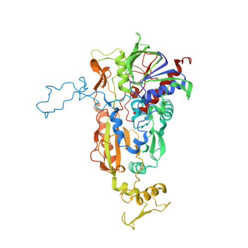
    Pyranose 2-oxidase catalyzes the oxidation of a number of carbohydrates using dioxygen; glucose, for example, is oxidized at carbon 2. The structure of pyranose 2-oxidase with the reaction product 2-keto-beta-d-glucose bound in the active center is reported in a new crystal form at 1.41 A resolution. The binding structure suggests that the alpha-anomer cannot be processed. The binding mode of the oxidized product was used to model other sugars accepted by the enzyme and to explain its specificity and catalytic rates. The reported structure at pH 6.0 shows a drastic conformational change in the loop of residues 454-461 (loop 454-461) at the active center compared to that of a closely homologous enzyme analyzed at pH 4.5 with a bound acetate inhibitor. In our structures, the loop is highly mobile and shifts to make way for the sugar to pass into the active center. Presumably, loop 454-461 functions as a gatekeeper. Apart from the wild-type enzyme, a thermostable variant was analyzed at 1.84 A resolution. In this variant, Glu542 is exchanged for a lysine. The observed stabilization could be a result of the mutated residue changing an ionic contact at a comparatively weak interface of the tetramer.


  • Organizational Affiliation

    Institut für Organische Chemie und Biochemie, Albert-Ludwigs-Universität, Albertstrasse 21, 79104 Freiburg im Breisgau, Germany.


Macromolecules
Find similar proteins by:  (by identity cutoff)  |  3D Structure
Entity ID: 1
MoleculeChains Sequence LengthOrganismDetailsImage
Pyranose 2-oxidase595Peniophora sp. SGMutation(s): 1 
Gene Names: p2oxpoxSG
EC: 1.1.3.10
UniProt
Find proteins for Q8J136 (Peniophora sp. (strain SG))
Explore Q8J136 
Go to UniProtKB:  Q8J136
Entity Groups  
Sequence Clusters30% Identity50% Identity70% Identity90% Identity95% Identity100% Identity
UniProt GroupQ8J136
Sequence Annotations
Expand
  • Reference Sequence
Small Molecules
Experimental Data & Validation

Experimental Data

  • Method: X-RAY DIFFRACTION
  • Resolution: 1.84 Å
  • R-Value Free: 0.186 
  • R-Value Work: 0.150 
  • R-Value Observed: 0.152 
  • Space Group: P 42 21 2
Unit Cell:
Length ( Å )Angle ( ˚ )
a = 101.931α = 90
b = 101.931β = 90
c = 118.602γ = 90
Software Package:
Software NamePurpose
REFMACrefinement
MAR345data collection
APRVdata scaling
EPMRphasing

Structure Validation

View Full Validation Report


Ligand Structure Quality Assessment 


Entry History 

Deposition Data

Revision History  (Full details and data files)

  • Version 1.0: 2006-06-13
    Type: Initial release
  • Version 1.1: 2008-05-01
    Changes: Version format compliance
  • Version 1.2: 2011-07-13
    Changes: Advisory, Derived calculations, Version format compliance
  • Version 1.3: 2017-10-18
    Changes: Refinement description
  • Version 1.4: 2021-10-20
    Changes: Data collection, Database references, Derived calculations