2F38

Crystal structure of prostaglandin F synathase containing bimatoprost


Experimental Data Snapshot

  • Method: X-RAY DIFFRACTION
  • Resolution: 2.00 Å
  • R-Value Free: 0.281 
  • R-Value Work: 0.220 
  • R-Value Observed: 0.221 

wwPDB Validation   3D Report Full Report


Ligand Structure Quality Assessment 


This is version 1.3 of the entry. See complete history


Literature

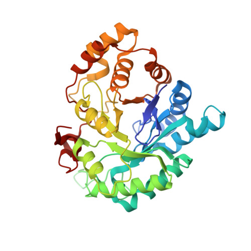
Prostaglandin F2alpha formation from prostaglandin H2 by prostaglandin F synthase (PGFS): crystal structure of PGFS containing bimatoprost.

Komoto, J.Yamada, T.Watanabe, K.Woodward, D.F.Takusagawa, F.

(2006) Biochemistry 45: 1987-1996

  • DOI: https://doi.org/10.1021/bi051861t
  • Primary Citation of Related Structures:  
    2F38

  • PubMed Abstract: 

    Prostaglandin H(2) (PGH(2)) formed from arachidonic acid is an unstable intermediate and is efficiently converted into more stable arachidonate metabolites by the action of enzymes. Prostaglandin F synthase (PGFS) has dual catalytic activities: formation of PGF(2)(alpha) from PGH(2) by the PGH(2) 9,11-endoperoxide reductase activity and 9alpha,11beta-PGF(2) (PGF(2)(alphabeta)) from PGD(2) by the PGD(2) 11-ketoreductase activity in the presence of NADPH. Bimatoprost (BMP), which is a highly effective ocular hypotensive agent, is a PGF(2)(alpha) analogue that inhibits both the PGD(2) 11-ketoreductase and PGH(2) 9,11-endoperoxide reductase activities of PGFS. To examine the catalytic mechanism of PGH(2) 9,11-endoperoxide reductase, a crystal structure of PGFS[NADPH + BMP] has been determined at 2.0 A resolution. BMP binds near the PGD(2) binding site, but the alpha- and omega-chains of BMP are locate on the omega- and alpha-chains of PGD(2), respectively. Consequently, the bound BMP and PGD(2) direct their opposite faces of the cyclopentane moieties toward the nicotinamide ring of the bound NADP. The alpha- and omega-chains of BMP are involved in H-bonding with protein residues, while the cyclopentane moiety is surrounded by water molecules and is not directly attached to either the protein or the bound NADPH, indicating that the cyclopentane moiety is movable in the active site. From the complex structure, two model structures of PGFS containing PGF(2)(alpha) and PGH(2) were built. On the basis of the model structures and inhibition data, a putative catalytic mechanism of PGH(2) 9,11-endoperoxide reductase of PGFS is proposed. Formation of PGF(2)(alpha) from PGH(2) most likely involves a direct hydride transfer from the bound NADPH to the endoperoxide of PGH(2) without the participation of specific amino acid residues.


  • Organizational Affiliation

    Department of Molecular Biosciences, University of Kansas, 1200 Sunnyside Avenue, Lawrence, Kansas 66045-7534, USA.


Macromolecules
Find similar proteins by:  (by identity cutoff)  |  3D Structure
Entity ID: 1
MoleculeChains Sequence LengthOrganismDetailsImage
Aldo-keto reductase family 1 member C3323Homo sapiensMutation(s): 0 
Gene Names: AKR1C3DDH1KIAA0119PGFS
EC: 1.3.1.20 (PDB Primary Data), 1.1.1.213 (PDB Primary Data), 1.1.1.188 (PDB Primary Data), 1.1.1.62 (PDB Primary Data)
UniProt & NIH Common Fund Data Resources
Find proteins for P42330 (Homo sapiens)
Explore P42330 
Go to UniProtKB:  P42330
PHAROS:  P42330
GTEx:  ENSG00000196139 
Entity Groups  
Sequence Clusters30% Identity50% Identity70% Identity90% Identity95% Identity100% Identity
UniProt GroupP42330
Sequence Annotations
Expand
  • Reference Sequence
Small Molecules
Ligands 2 Unique
IDChains Name / Formula / InChI Key2D Diagram3D Interactions
NAP
Query on NAP

Download Ideal Coordinates CCD File 
B [auth A]NADP NICOTINAMIDE-ADENINE-DINUCLEOTIDE PHOSPHATE
C21 H28 N7 O17 P3
XJLXINKUBYWONI-NNYOXOHSSA-N
15M
Query on 15M

Download Ideal Coordinates CCD File 
C [auth A](5Z)-7-{(1R,2R,3R,5S)-3,5-DIHYDROXY-2-[(1E,3S)-3-HYDROXY-5-PHENYLPENT-1-ENYL]CYCLOPENTYL}-N-ETHYLHEPT-5-ENAMIDE
C25 H37 N O4
AQOKCDNYWBIDND-FTOWTWDKSA-N
Binding Affinity Annotations 
IDSourceBinding Affinity
15M Binding MOAD:  2F38 IC50: 6000 (nM) from 1 assay(s)
BindingDB:  2F38 IC50: 5000 (nM) from 1 assay(s)
Experimental Data & Validation

Experimental Data

  • Method: X-RAY DIFFRACTION
  • Resolution: 2.00 Å
  • R-Value Free: 0.281 
  • R-Value Work: 0.220 
  • R-Value Observed: 0.221 
  • Space Group: P 1 21 1
Unit Cell:
Length ( Å )Angle ( ˚ )
a = 44.27α = 90
b = 78.16β = 100.2
c = 48.89γ = 90
Software Package:
Software NamePurpose
DENZOdata reduction
SCALEPACKdata scaling
X-PLORmodel building
X-PLORrefinement
X-PLORphasing

Structure Validation

View Full Validation Report


Ligand Structure Quality Assessment 


Entry History 

Deposition Data

Revision History  (Full details and data files)

  • Version 1.0: 2006-10-31
    Type: Initial release
  • Version 1.1: 2008-05-01
    Changes: Version format compliance
  • Version 1.2: 2011-07-13
    Changes: Version format compliance
  • Version 1.3: 2023-08-23
    Changes: Data collection, Database references, Derived calculations, Refinement description