2F31

Crystal structure of the autoinhibitory switch in Formin mDia1; the DID/DAD complex


Experimental Data Snapshot

  • Method: X-RAY DIFFRACTION
  • Resolution: 2.10 Å
  • R-Value Free: 0.312 
  • R-Value Work: 0.229 
  • R-Value Observed: 0.233 

wwPDB Validation   3D Report Full Report


This is version 1.4 of the entry. See complete history


Literature

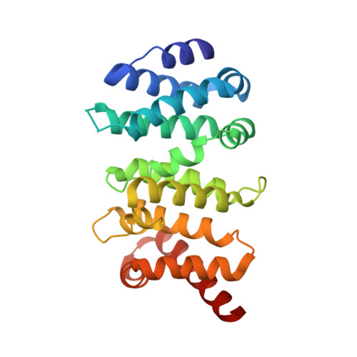
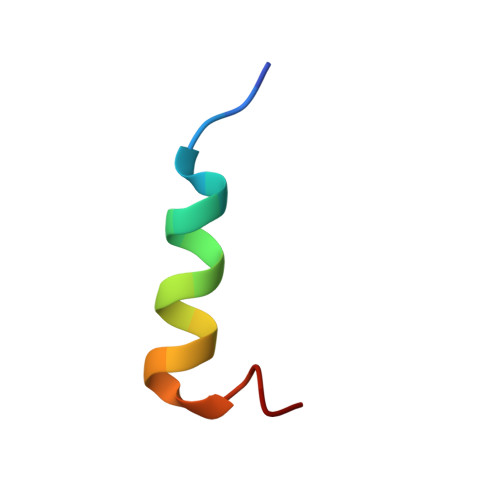
Structure of the Autoinhibitory Switch in Formin mDia1

Nezami, A.G.Poy, F.Eck, M.J.

(2006) Structure 14: 257-263

  • DOI: https://doi.org/10.1016/j.str.2005.12.003
  • Primary Citation of Related Structures:  
    2F31

  • PubMed Abstract: 

    Diaphanous-related formins (DRFs) regulate the nucleation and polymerization of unbranched actin filaments. The activity of DRFs is inhibited by an intramolecular interaction between their N-terminal regulatory region and a conserved C-terminal segment termed the Diaphanous autoinhibitory domain (DAD). Binding of GTP bound Rho to the mDia1 N terminus releases this autoinhibitory restraint. Here, we describe the crystal structure of the DAD segment of mDia1 in complex with the relevant N-terminal fragment, termed the DID domain. The structure reveals that the DAD segment forms an amphipathic helix that binds a conserved, concave surface on the DID domain. Comparison with the structure of the mDia1 N terminus bound to RhoC suggests that release of the autoinhibitory DAD interaction is accomplished largely by Rho-induced restructuring of the adjacent GTPase binding subdomain (GBD), but also by electrostatic repulsion and a small, direct steric occlusion of the DAD binding cleft by Rho itself.


  • Organizational Affiliation

    Department of Biological Chemistry and Molecular Pharmacology, Harvard Medical School, 44 Binney Street, Boston, Massachusetts 02115, USA.


Macromolecules
Find similar proteins by:  (by identity cutoff)  |  3D Structure
Entity ID: 1
MoleculeChains Sequence LengthOrganismDetailsImage
Diaphanous protein homolog 1233Mus musculusMutation(s): 0 
Gene Names: Diaph1Diap1
UniProt & NIH Common Fund Data Resources
Find proteins for Q3US76 (Mus musculus)
Explore Q3US76 
Go to UniProtKB:  Q3US76
IMPC:  MGI:1194490
Entity Groups  
Sequence Clusters30% Identity50% Identity70% Identity90% Identity95% Identity100% Identity
UniProt GroupQ3US76
Sequence Annotations
Expand
  • Reference Sequence

Find similar proteins by:  Sequence   |   3D Structure  

Entity ID: 2
MoleculeChains Sequence LengthOrganismDetailsImage
Diaphanous protein homolog 120Mus musculusMutation(s): 0 
Gene Names: Diaph1Diap1
UniProt & NIH Common Fund Data Resources
Find proteins for O08808 (Mus musculus)
Explore O08808 
Go to UniProtKB:  O08808
IMPC:  MGI:1194490
Entity Groups  
Sequence Clusters30% Identity50% Identity70% Identity90% Identity95% Identity100% Identity
UniProt GroupO08808
Sequence Annotations
Expand
  • Reference Sequence
Experimental Data & Validation

Experimental Data

  • Method: X-RAY DIFFRACTION
  • Resolution: 2.10 Å
  • R-Value Free: 0.312 
  • R-Value Work: 0.229 
  • R-Value Observed: 0.233 
  • Space Group: C 1 2 1
Unit Cell:
Length ( Å )Angle ( ˚ )
a = 102.16α = 90
b = 65.79β = 104.08
c = 46.86γ = 90
Software Package:
Software NamePurpose
XSCALEdata scaling
PHASERphasing
REFMACrefinement
PDB_EXTRACTdata extraction
MAR345data collection
XDSdata scaling

Structure Validation

View Full Validation Report


Entry History 

Deposition Data

Revision History  (Full details and data files)

  • Version 1.0: 2006-05-30
    Type: Initial release
  • Version 1.1: 2008-05-01
    Changes: Version format compliance
  • Version 1.2: 2011-07-13
    Changes: Version format compliance
  • Version 1.3: 2017-10-18
    Changes: Refinement description
  • Version 1.4: 2024-02-14
    Changes: Data collection, Database references