2F1B | pdb_00002f1b

GOLGI ALPHA-MANNOSIDASE II COMPLEX WITH (2R,3R,4S,5R)-2-({[(1R)-2-hydroxy-1-phenylethyl]amino}methyl)-5-methylpyrrolidine-3,4-diol


Experimental Data Snapshot

  • Method: X-RAY DIFFRACTION
  • Resolution: 1.45 Å
  • R-Value Free: 
    0.194 (Depositor), 0.196 (DCC) 
  • R-Value Work: 
    0.173 (Depositor), 0.176 (DCC) 
  • R-Value Observed: 
    0.173 (Depositor) 

Starting Model: experimental
View more details

wwPDB Validation 3D Report Full Report

Validation slider image for 2F1B

Ligand Structure Quality Assessment 


This is version 1.5 of the entry. See complete history

Literature

Evaluation of docking programs for predicting binding of Golgi alpha-mannosidase II inhibitors: a comparison with crystallography.

Englebienne, P.Fiaux, H.Kuntz, D.A.Corbeil, C.R.Gerber-Lemaire, S.Rose, D.R.Moitessier, N.

(2007) Proteins 69: 160-176

  • DOI: https://doi.org/10.1002/prot.21479
  • Primary Citation Related Structures: 
    2F18, 2F1A, 2F1B

  • PubMed Abstract: 

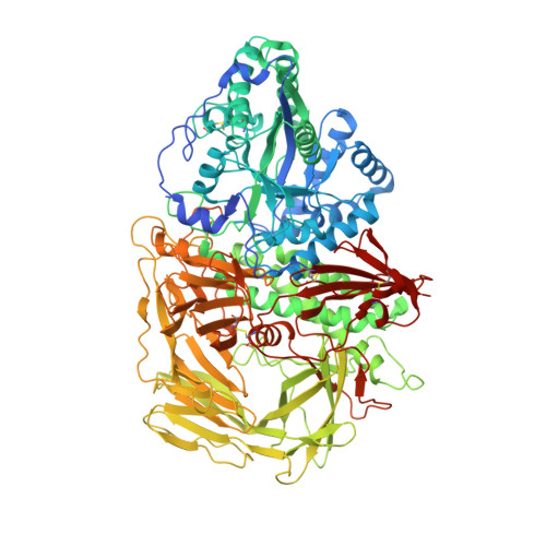
    Golgi alpha-mannosidase II (GMII), a zinc-dependent glycosyl hydrolase, is a promising target for drug development in anti-tumor therapies. Using X-ray crystallography, we have determined the structure of Drosophila melanogaster GMII (dGMII) complexed with three different inhibitors exhibiting IC50's ranging from 80 to 1000 microM. These structures, along with those of seven other available dGMII/inhibitor complexes, were then used as a basis for the evaluation of seven docking programs (GOLD, Glide, FlexX, AutoDock, eHiTS, LigandFit, and FITTED). We found that small inhibitors could be accurately docked by most of the software, while docking of larger compounds (i.e., those with extended aromatic cycles or long aliphatic chains) was more problematic. Overall, Glide provided the best docking results, with the most accurately predicted binding around the active site zinc atom. Further evaluation of Glide's performance revealed its ability to extract active compounds from a benchmark library of decoys.


  • Organizational Affiliation
    • Department of Chemistry, McGill University, Montréal, Québec H3A 2K6, Canada.

Macromolecule Content 

  • Total Structure Weight: 120.37 kDa 
  • Atom Count: 9,491 
  • Modeled Residue Count: 1,014 
  • Deposited Residue Count: 1,045 
  • Unique protein chains: 1

Macromolecules

Find similar proteins by:|  3D Structure
Entity ID: 1
MoleculeChains  Sequence LengthOrganismDetailsImage
alpha-mannosidase II1,045Drosophila melanogasterMutation(s): 0 
Gene Names: alpha-Man-IIGmII
EC: 3.2.1.114
UniProt
Find proteins for Q24451 (Drosophila melanogaster)
Explore Q24451 
Go to UniProtKB:  Q24451
Entity Groups
Sequence Clusters30% Identity50% Identity70% Identity90% Identity95% Identity100% Identity
UniProt GroupQ24451
Glycosylation
Glycosylation Sites: 1Go to GlyGen: Q24451-1
Sequence Annotations
Expand
Reference Sequence

Small Molecules

Experimental Data & Validation

Experimental Data

  • Method: X-RAY DIFFRACTION
  • Resolution: 1.45 Å
  • R-Value Free:  0.194 (Depositor), 0.196 (DCC) 
  • R-Value Work:  0.173 (Depositor), 0.176 (DCC) 
  • R-Value Observed: 0.173 (Depositor) 
Space Group: P 21 21 21
Unit Cell:
Length ( Å )Angle ( ˚ )
a = 69.433α = 90
b = 110.578β = 90
c = 139.89γ = 90
Software Package:
Software NamePurpose
CNSrefinement
DENZOdata reduction
SCALEPACKdata scaling
CNSphasing

Structure Validation

View Full Validation Report


Ligand Structure Quality Assessment 


Entry History 

Deposition Data

Revision History  (Full details and data files)

  • Version 1.0: 2006-12-05
    Type: Initial release
  • Version 1.1: 2007-10-16
    Changes: Version format compliance
  • Version 1.2: 2011-07-13
    Changes: Non-polymer description, Version format compliance
  • Version 1.3: 2020-07-29
    Type: Remediation
    Reason: Carbohydrate remediation
    Changes: Advisory, Data collection, Database references, Derived calculations, Structure summary
  • Version 1.4: 2023-08-23
    Changes: Data collection, Database references, Refinement description, Structure summary
  • Version 1.5: 2024-11-20
    Changes: Structure summary