2F08 | pdb_00002f08

Crystal structure of a major house dust mite allergen, Derf 2


Experimental Data Snapshot

  • Method: X-RAY DIFFRACTION
  • Resolution: 2.20 Å
  • R-Value Free: 
    0.245 (Depositor), 0.244 (DCC) 
  • R-Value Work: 
    0.206 (Depositor), 0.207 (DCC) 
  • R-Value Observed: 
    0.206 (Depositor) 

wwPDB Validation 3D Report Full Report

Validation slider image for 2F08

Ligand Structure Quality Assessment 


This is version 1.3 of the entry. See complete history

Literature

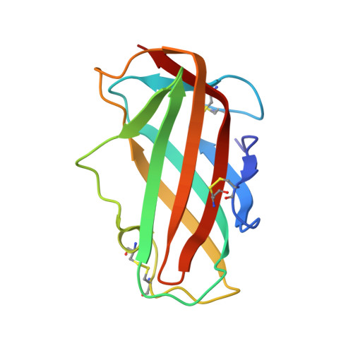
Crystal structure and some properties of a major house dust mite allergen, Derf 2

Suzuki, M.Tanaka, Y.Korematsu, S.Mikami, B.Minato, N.

(2006) Biochem Biophys Res Commun 339: 679-686

  • DOI: https://doi.org/10.1016/j.bbrc.2005.11.065
  • Primary Citation Related Structures: 
    2F08

  • PubMed Abstract: 

    Pyroglyphid house dust mites are a major source of allergens in house dust. Mite allergens sensitize and induce asthma, rhinitis, and eczema in a large portion of patients with allergic diseases. Here, the crystal structure of a major mite allergen, Derf 2, derived from Dermatophagoides farinae was solved by single isomorphous replacement method with anomalous scattering (SIRAS) at 2.1A resolution. The present study also demonstrated that the conformation of the allergen was critical in the determination of Th1/Th2 shift based on physicochemical and immunological analyses. This indicates that rigidly folded and singly dispersed structure is essentially required for the generation of Th2 type cells by the allergen, while conformational variant protein leads to Th1 skewing, irrespective of the same amino acid sequence. This structure/function relationship may allow us to develop a novel strategy for hyposensitization therapy in patients with allergic diseases triggered by house dust mite allergens.


  • Organizational Affiliation
    • Laboratory of Immunology and Cell Biology, Graduate School of Biostudies, Kyoto University, Yoshidakonoe-Cho, Sakyo-Ku, Kyoto 606-8501, Japan.

Macromolecule Content 

  • Total Structure Weight: 56.58 kDa 
  • Atom Count: 4,120 
  • Modeled Residue Count: 516 
  • Deposited Residue Count: 516 
  • Unique protein chains: 1

Macromolecules

Find similar proteins by:|  3D Structure
Entity ID: 1
MoleculeChains  Sequence LengthOrganismDetailsImage
mite allergen Der f II
A, B, C, D
129Dermatophagoides farinaeMutation(s): 0 
UniProt
Find proteins for Q00855 (Dermatophagoides farinae)
Explore Q00855 
Go to UniProtKB:  Q00855
Entity Groups
Sequence Clusters30% Identity50% Identity70% Identity90% Identity95% Identity100% Identity
UniProt GroupQ00855
Sequence Annotations
Expand
Reference Sequence

Small Molecules

Ligands 1 Unique
IDChains Name / Formula / InChI Key2D Diagram3D Interactions
P4C

Query on P4C


Download:Ideal Coordinates CCD File
E [auth C]O-ACETALDEHYDYL-HEXAETHYLENE GLYCOL
C14 H28 O8
CTLLATPOKUEFSQ-UHFFFAOYSA-N

Experimental Data & Validation

Experimental Data

  • Method: X-RAY DIFFRACTION
  • Resolution: 2.20 Å
  • R-Value Free:  0.245 (Depositor), 0.244 (DCC) 
  • R-Value Work:  0.206 (Depositor), 0.207 (DCC) 
  • R-Value Observed: 0.206 (Depositor) 
Space Group: C 1 2 1
Unit Cell:
Length ( Å )Angle ( ˚ )
a = 106.124α = 90
b = 54.328β = 91.35
c = 102.354γ = 90
Software Package:
Software NamePurpose
CNSrefinement
SAINTdata reduction
SAINTdata scaling
PHASESphasing

Structure Validation

View Full Validation Report


Ligand Structure Quality Assessment 


Entry History 

Deposition Data

Revision History  (Full details and data files)

  • Version 1.0: 2005-11-29
    Type: Initial release
  • Version 1.1: 2008-05-01
    Changes: Version format compliance
  • Version 1.2: 2011-07-13
    Changes: Version format compliance
  • Version 1.3: 2024-11-13
    Changes: Data collection, Database references, Derived calculations, Structure summary