2EWF

Crystal structure of the site-specific DNA nickase N.BspD6I


Experimental Data Snapshot

  • Method: X-RAY DIFFRACTION
  • Resolution: 1.84 Å
  • R-Value Free: 0.236 
  • R-Value Work: 0.183 
  • R-Value Observed: 0.200 

wwPDB Validation   3D Report Full Report


This is version 1.3 of the entry. See complete history


Literature

Structural analysis of the heterodimeric type IIS restriction endonuclease R.BspD6I acting as a complex between a monomeric site-specific nickase and a catalytic subunit.

Kachalova, G.S.Rogulin, E.A.Yunusova, A.K.Artyukh, R.I.Perevyazova, T.A.Matvienko, N.I.Zheleznaya, L.A.Bartunik, H.D.

(2008) J Mol Biol 384: 489-502

  • DOI: https://doi.org/10.1016/j.jmb.2008.09.033
  • Primary Citation of Related Structures:  
    2EWF, 2P14

  • PubMed Abstract: 

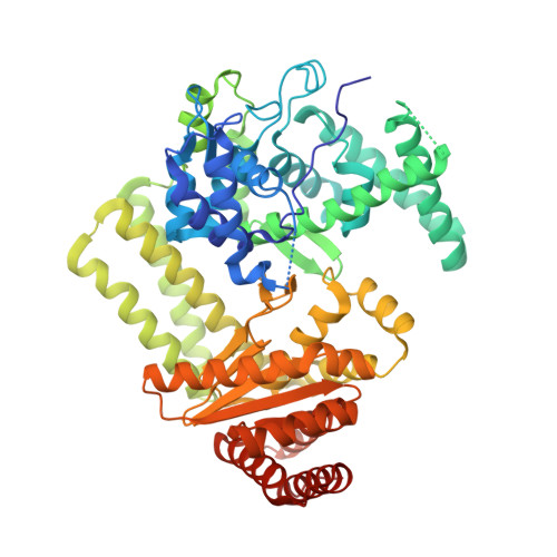
    The heterodimeric restriction endonuclease R.BspD6I from Bacillus species D6 recognizes a pseudosymmetric sequence and cuts both DNA strands outside the recognition sequence. The large subunit, Nt.BspD6I, acts as a type IIS site-specific monomeric nicking endonuclease. The isolated small subunit, ss.BspD6I, does not bind DNA and is not catalytically active. We solved the crystal structures of Nt.BspD6I and ss.BspD6I at high resolution. Nt.BspD6I consists of three domains, two of which exhibit structural similarity to the recognition and cleavage domains of FokI. ss.BspD6I has a fold similar to that of the cleavage domain of Nt.BspD6I, each containing a PD-(D/E)XK motif and a histidine as an additional putative catalytic residue. In contrast to the DNA-bound FokI structure, in which the cleavage domain is rotated away from the DNA, the crystal structure of Nt.BspD6I shows the recognition and cleavage domains in favorable orientations for interactions with DNA. Docking models of complexes of Nt.BspD6I and R.BspD6I with cognate DNA were constructed on the basis of structural similarity to individual domains of FokI, R.BpuJI and HindIII. A three-helix bundle forming an interdomain linker in Nt.BspD6I acts as a rigid spacer adjusting the orientations of the spatially separated domains to match the distance between the recognition and cleavage sites accurately.


  • Organizational Affiliation

    Max Planck Unit for Structural Molecular Biology, MPG-ASMB c/o DESY, Notkestrasse 85, 22603 Hamburg, Germany.


Macromolecules
Find similar proteins by:  (by identity cutoff)  |  3D Structure
Entity ID: 1
MoleculeChains Sequence LengthOrganismDetailsImage
Nicking endonuclease N.BspD6I610Bacillus sp. D6Mutation(s): 0 
UniProt
Find proteins for A3FEV7 (Bacillus sp. D6)
Explore A3FEV7 
Go to UniProtKB:  A3FEV7
Entity Groups  
Sequence Clusters30% Identity50% Identity70% Identity90% Identity95% Identity100% Identity
UniProt GroupA3FEV7
Sequence Annotations
Expand
  • Reference Sequence
Small Molecules
Ligands 1 Unique
IDChains Name / Formula / InChI Key2D Diagram3D Interactions
BR
Query on BR

Download Ideal Coordinates CCD File 
B [auth A]
C [auth A]
D [auth A]
E [auth A]
F [auth A]
B [auth A],
C [auth A],
D [auth A],
E [auth A],
F [auth A],
G [auth A],
H [auth A],
I [auth A],
J [auth A],
K [auth A],
L [auth A],
M [auth A],
N [auth A]
BROMIDE ION
Br
CPELXLSAUQHCOX-UHFFFAOYSA-M
Experimental Data & Validation

Experimental Data

  • Method: X-RAY DIFFRACTION
  • Resolution: 1.84 Å
  • R-Value Free: 0.236 
  • R-Value Work: 0.183 
  • R-Value Observed: 0.200 
  • Space Group: P 1 21 1
Unit Cell:
Length ( Å )Angle ( ˚ )
a = 59.553α = 90
b = 92.562β = 111.88
c = 76.349γ = 90
Software Package:
Software NamePurpose
SHELXmodel building
SHELXL-97refinement
DENZOdata reduction
SCALEPACKdata scaling
SHELXSphasing

Structure Validation

View Full Validation Report


Entry History 

Deposition Data

Revision History  (Full details and data files)

  • Version 1.0: 2006-11-21
    Type: Initial release
  • Version 1.1: 2008-05-01
    Changes: Version format compliance
  • Version 1.2: 2011-07-13
    Changes: Source and taxonomy, Version format compliance
  • Version 1.3: 2024-02-14
    Changes: Data collection, Database references, Derived calculations