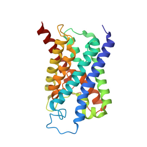
2EVU

Crystal structure of aquaporin AqpM at 2.3A resolution


Experimental Data Snapshot

  • Method: X-RAY DIFFRACTION
  • Resolution: 2.30 Å
  • R-Value Free: 0.226 
  • R-Value Work: 0.188 
  • R-Value Observed: 0.188 

wwPDB Validation   3D Report Full Report


Ligand Structure Quality Assessment 


This is version 1.4 of the entry. See complete history


Literature

Structural basis for conductance by the archaeal aquaporin AqpM at 1.68 A.

Lee, J.K.Kozono, D.Remis, J.Kitagawa, Y.Agre, P.Stroud, R.M.

(2005) Proc Natl Acad Sci U S A 102: 18932-18937

  • DOI: https://doi.org/10.1073/pnas.0509469102
  • Primary Citation of Related Structures:  
    2EVU, 2F2B

  • PubMed Abstract: 

    To explore the structural basis of the unique selectivity spectrum and conductance of the transmembrane channel protein AqpM from the archaeon Methanothermobacter marburgensis, we determined the structure of AqpM to 1.68-A resolution by x-ray crystallography. The structure establishes AqpM as being in a unique subdivision between the two major subdivisions of aquaporins, the water-selective aquaporins, and the water-plus-glycerol-conducting aquaglyceroporins. In AqpM, isoleucine replaces a key histidine residue found in the lumen of water channels, which becomes a glycine residue in aquaglyceroporins. As a result of this and other side-chain substituents in the walls of the channel, the channel is intermediate in size and exhibits differentially tuned electrostatics when compared with the other subfamilies.


  • Organizational Affiliation

    Macromolecular Structure Group, Department of Biochemistry and Biophysics, University of California, S-412C Genentech Hall, 600 16th Street, San Francisco, CA 94143-2240, USA.


Macromolecules
Find similar proteins by:  (by identity cutoff)  |  3D Structure
Entity ID: 1
MoleculeChains Sequence LengthOrganismDetailsImage
Aquaporin aqpM246Methanothermobacter marburgensis str. MarburgMutation(s): 0 
Gene Names: aqpM
Membrane Entity: Yes 
UniProt
Find proteins for Q9C4Z5 (Methanothermobacter marburgensis (strain ATCC BAA-927 / DSM 2133 / JCM 14651 / NBRC 100331 / OCM 82 / Marburg))
Explore Q9C4Z5 
Go to UniProtKB:  Q9C4Z5
Entity Groups  
Sequence Clusters30% Identity50% Identity70% Identity90% Identity95% Identity100% Identity
UniProt GroupQ9C4Z5
Sequence Annotations
Expand
  • Reference Sequence
Small Molecules
Ligands 2 Unique
IDChains Name / Formula / InChI Key2D Diagram3D Interactions
BOG
Query on BOG

Download Ideal Coordinates CCD File 
B [auth A],
C [auth A]
octyl beta-D-glucopyranoside
C14 H28 O6
HEGSGKPQLMEBJL-RKQHYHRCSA-N
GOL
Query on GOL

Download Ideal Coordinates CCD File 
D [auth A]
E [auth A]
F [auth A]
G [auth A]
H [auth A]
D [auth A],
E [auth A],
F [auth A],
G [auth A],
H [auth A],
I [auth A]
GLYCEROL
C3 H8 O3
PEDCQBHIVMGVHV-UHFFFAOYSA-N
Experimental Data & Validation

Experimental Data

  • Method: X-RAY DIFFRACTION
  • Resolution: 2.30 Å
  • R-Value Free: 0.226 
  • R-Value Work: 0.188 
  • R-Value Observed: 0.188 
  • Space Group: I 4
Unit Cell:
Length ( Å )Angle ( ˚ )
a = 93.887α = 90
b = 93.887β = 90
c = 77.744γ = 90
Software Package:
Software NamePurpose
DENZOdata reduction
SCALEPACKdata scaling
CNSrefinement
CNSphasing

Structure Validation

View Full Validation Report


Ligand Structure Quality Assessment 


Entry History 

Deposition Data

Revision History  (Full details and data files)

  • Version 1.0: 2005-12-06
    Type: Initial release
  • Version 1.1: 2008-05-01
    Changes: Version format compliance
  • Version 1.2: 2011-07-13
    Changes: Derived calculations, Version format compliance
  • Version 1.3: 2020-07-29
    Type: Remediation
    Reason: Carbohydrate remediation
    Changes: Data collection, Derived calculations, Structure summary
  • Version 1.4: 2024-02-14
    Changes: Data collection, Database references, Structure summary