2EV8 | pdb_00002ev8

Solution structure of the erythroid p55 PDZ domain


Experimental Data Snapshot

  • Method: SOLUTION NMR
  • Conformers Calculated: 100 
  • Conformers Submitted: 20 
  • Selection Criteria: structures with the lowest energy 

wwPDB Validation 3D Report Full Report

Validation slider image for 2EV8

This is version 1.4 of the entry. See complete history

Literature

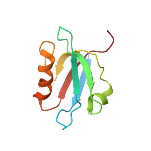
Solution structure of human erythroid p55 PDZ domain

Kusunoki, H.Kohno, T.

(2006) Proteins 64: 804-807

Macromolecule Content 

  • Total Structure Weight: 10.95 kDa 
  • Atom Count: 663 
  • Modeled Residue Count: 85 
  • Deposited Residue Count: 97 
  • Unique protein chains: 1

Macromolecules

Find similar proteins by:|  3D Structure
Entity ID: 1
MoleculeChains  Sequence LengthOrganismDetailsImage
55 kDa erythrocyte membrane protein97Homo sapiensMutation(s): 0 
UniProt & NIH Common Fund Data Resources
Find proteins for Q00013 (Homo sapiens)
Explore Q00013 
Go to UniProtKB:  Q00013
PHAROS:  Q00013
GTEx:  ENSG00000130830 
Entity Groups
Sequence Clusters30% Identity50% Identity70% Identity90% Identity95% Identity100% Identity
UniProt GroupQ00013
Sequence Annotations
Expand
Reference Sequence

Experimental Data & Validation

Experimental Data

  • Method: SOLUTION NMR
  • Conformers Calculated: 100 
  • Conformers Submitted: 20 
  • Selection Criteria: structures with the lowest energy 

Structure Validation

View Full Validation Report


Entry History 

Deposition Data

Revision History  (Full details and data files)

  • Version 1.0: 2006-10-10
    Type: Initial release
  • Version 1.1: 2008-05-01
    Changes: Version format compliance
  • Version 1.2: 2011-07-13
    Changes: Version format compliance
  • Version 1.3: 2022-03-09
    Changes: Data collection, Database references, Derived calculations
  • Version 1.4: 2024-05-29
    Changes: Data collection