2ERS | pdb_00002ers

Solution structure of the Interleukin-15 receptor sushi domain


Experimental Data Snapshot

  • Method: SOLUTION NMR
  • Conformers Calculated: 20 
  • Conformers Submitted: 
  • Selection Criteria: structures with the lowest energy 

wwPDB Validation 3D Report Full Report

Validation slider image for 2ERS

This is version 1.4 of the entry. See complete history

Literature

The structure of the interleukin-15 alpha receptor and its implications for ligand binding.

Lorenzen, I.Dingley, A.J.Jacques, Y.Grotzinger, J.

(2006) J Biological Chem 281: 6642-6647

  • DOI: https://doi.org/10.1074/jbc.M513118200
  • Primary Citation Related Structures: 
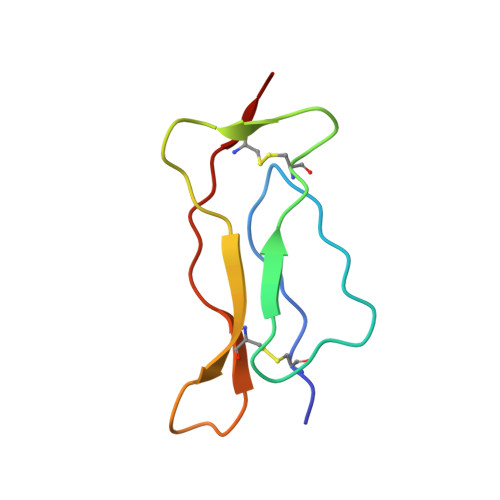
    2ERS

  • PubMed Abstract: 

    Interleukin (IL)-15 is a member of the small four alpha-helix bundle family of cytokines. IL-15 was discovered by its ability to mimic IL-2-mediated T-cell proliferation. Both cytokines share the beta and gamma receptor chains of the IL-2 receptor for signal transduction. However, in addition, they target specific alpha chain receptors IL-15Ralpha and IL-2Ralpha, respectively. The exceptionally high affinity binding of IL-15 to IL-15Ralpha is mediated by its sushi domain. Here we present the solution structure of the IL-15Ralpha sushi domain solved by NMR spectroscopy and a model of its complex with IL-15. The model shows that, rather than the familiar hydrophobic forces dominating the interaction interface between cytokines and their cognate receptors, the interaction between the IL-15 and IL-15Ralpha complex involves a large network of ionic interactions. This type of interaction explains the exceptionally high affinity of the IL-15.IL-15Ralpha complex, which is essential for the biological effects of this important cytokine and which is not observed in other cytokine/cytokine receptor complexes.


  • Organizational Affiliation
    • Biochemisches Institut der Christian-Albrechts-Universität Kiel, Olshausenstrasse 40, 24118 Kiel, Germany.

Macromolecule Content 

  • Total Structure Weight: 7.47 kDa 
  • Atom Count: 520 
  • Modeled Residue Count: 66 
  • Deposited Residue Count: 66 
  • Unique protein chains: 1

Macromolecules

Find similar proteins by:|  3D Structure
Entity ID: 1
MoleculeChains  Sequence LengthOrganismDetailsImage
Interleukin-15 receptor alpha chain66Homo sapiensMutation(s): 0 
UniProt & NIH Common Fund Data Resources
Find proteins for Q13261 (Homo sapiens)
Explore Q13261 
Go to UniProtKB:  Q13261
PHAROS:  Q13261
GTEx:  ENSG00000134470 
Entity Groups
Sequence Clusters30% Identity50% Identity70% Identity90% Identity95% Identity100% Identity
UniProt GroupQ13261
Sequence Annotations
Expand
Reference Sequence

Experimental Data & Validation

Experimental Data

  • Method: SOLUTION NMR
  • Conformers Calculated: 20 
  • Conformers Submitted: 
  • Selection Criteria: structures with the lowest energy 

Structure Validation

View Full Validation Report


Entry History 

Deposition Data

Revision History  (Full details and data files)

  • Version 1.0: 2006-02-07
    Type: Initial release
  • Version 1.1: 2008-05-01
    Changes: Version format compliance
  • Version 1.2: 2011-07-13
    Changes: Version format compliance
  • Version 1.3: 2022-03-09
    Changes: Database references, Derived calculations
  • Version 1.4: 2024-10-16
    Changes: Data collection, Structure summary