2ELG | pdb_00002elg

The rare crystallographic structure of d(CGCGCG)2: The natural spermidine molecule bound to the minor groove of left-handed Z-DNA d(CGCGCG)2 at 10 degree celsius


Experimental Data Snapshot

  • Method: X-RAY DIFFRACTION
  • Resolution: 1.00 Å
  • R-Value Free: 
    0.247 (Depositor) 
  • R-Value Work: 
    0.232 (Depositor), 0.229 (DCC) 
  • R-Value Observed: 
    0.244 (Depositor) 

Starting Model: experimental
View more details

wwPDB Validation 3D Report Full Report

Validation slider image for 2ELG

This is version 1.2 of the entry. See complete history

Literature

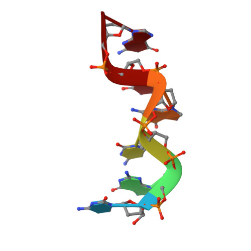
The rare crystallographic structure of d(CGCGCG)(2): The natural spermidine molecule bound to the minor groove of left-handed Z-DNA d(CGCGCG)(2) at 10 degrees C

Ohishi, H.Tozuka, Y.Da-Yang, Z.Ishida, T.Nakatani, K.

(2007) Biochem Biophys Res Commun 358: 24-28

  • DOI: https://doi.org/10.1016/j.bbrc.2007.04.026
  • Primary Citation Related Structures: 
    2ELG

  • PubMed Abstract: 

    Several crystal structure analyses of complexes of synthetic polyamine compounds, including N(1)-(2-(2-aminoethylamino))ethyl)ethane-1,2-diamine PA(222) and N(1)-(2-(2-(2-aminoethylamino)ethylamino)ethyl)ethane-1,2-diamine PA(2222), and left-handed Z-DNA d(CGCGCG)(2) have been reported. However, until now, there have been no examples of naturally occurring polyamines bound to the minor groove of the left-handed Z-DNA of d(CGCGCG)(2) molecule. We have found that spermidine, a natural polyamine, is connected to the minor groove of left-handed Z-DNA of d(CGCGCG)(2) molecule in a crystalline complex grown at 10 degrees C. The electron density of the DNA molecule was clear enough to determine that the spermidine was connected in the minor groove of two symmetry related molecules of left-handed Z-DNA d(CGCGCG)(2). This is the first example that a spermidine molecule can form a bridge conformation between two symmetry related molecules of left-handed Z-DNA d(CGCGCG)(2) in the minor groove.


  • Organizational Affiliation
    • Osaka University of Pharmaceutical Sciences, 4-20-1, Nasahara, Takatsuki, Osaka 569-1094, Japan. ohishi@gly.oups.ac.jp

Macromolecule Content 

  • Total Structure Weight: 3.86 kDa 
  • Atom Count: 321 
  • Modeled Residue Count: 12 
  • Deposited Residue Count: 12 
  • Unique nucleic acid chains: 1

Macromolecules

Find similar nucleic acids by:  Sequence
Entity ID: 1
MoleculeChains LengthOrganismImage
DNA (5'-D(*DCP*DGP*DCP*DGP*DCP*DG)-3')
A, B
6N/A
Sequence Annotations
Expand
Reference Sequence

Experimental Data & Validation

Experimental Data

  • Method: X-RAY DIFFRACTION
  • Resolution: 1.00 Å
  • R-Value Free:  0.247 (Depositor) 
  • R-Value Work:  0.232 (Depositor), 0.229 (DCC) 
  • R-Value Observed: 0.244 (Depositor) 
Space Group: P 21 21 21
Unit Cell:
Length ( Å )Angle ( ˚ )
a = 17.847α = 90
b = 30.988β = 90
c = 44.022γ = 90
Software Package:
Software NamePurpose
ADSCdata collection
MERLOTphasing
X-PLORrefinement
MOSFLMdata reduction
SCALAdata scaling

Structure Validation

View Full Validation Report


Entry History 

Deposition Data

Revision History  (Full details and data files)

  • Version 1.0: 2008-04-01
    Type: Initial release
  • Version 1.1: 2011-07-13
    Changes: Version format compliance
  • Version 1.2: 2023-10-25
    Changes: Data collection, Database references, Derived calculations, Refinement description