2EJY | pdb_00002ejy

Solution structure of the p55 PDZ T85C domain complexed with the glycophorin C F127C peptide


Experimental Data Snapshot

  • Method: SOLUTION NMR
  • Conformers Calculated: 100 
  • Conformers Submitted: 20 
  • Selection Criteria: structures with the lowest energy 

wwPDB Validation 3D Report Full Report

Validation slider image for 2EJY

This is version 1.3 of the entry. See complete history

Literature

Structural insight into the interaction between the p55 PDZ domain and glycophorin C

Kusunoki, H.Kohno, T.

(2007) Biochem Biophys Res Commun 359: 972-978

  • DOI: https://doi.org/10.1016/j.bbrc.2007.05.215
  • Primary Citation Related Structures: 
    2EJY

  • PubMed Abstract: 

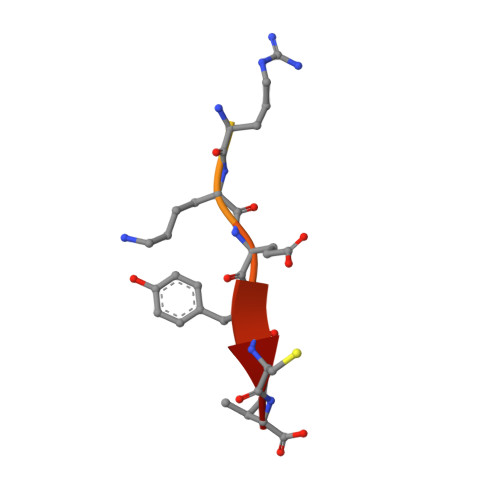
    p55, a member of the membrane-associated guanylate kinase family, includes a PDZ domain that specifically interacts with the C-terminal region of glycophorin C in the ternary complex of p55, protein 4.1 and glycophorin C. Here we present the first NMR-derived complex structure of the p55 PDZ domain and the C-terminal peptide of glycophorin C, obtained by using a threonine to cysteine (T85C) mutant of the p55 PDZ domain and a phenylalanine to cysteine (F127C) mutant of the glycophorin C peptide. Our NMR results revealed that the two designed mutant molecules retain the specific interaction manner that exists between the wild type molecules and can facilitate the structure determination by NMR, due to the stable complex formation via an intermolecular disulfide bond. The complex structure provides insight into the specific interaction of the p55 PDZ domain with the two key residues, Ile128 and Tyr126, of glycophorin C.


  • Organizational Affiliation
    • Mitsubishi Kagaku Institute of Life Sciences (MITILS), 11 Minamiooya, Machida, Tokyo 194-8511, Japan.

Macromolecule Content 

  • Total Structure Weight: 12.3 kDa 
  • Atom Count: 718 
  • Modeled Residue Count: 91 
  • Deposited Residue Count: 109 
  • Unique protein chains: 2

Macromolecules

Find similar proteins by:|  3D Structure
Entity ID: 1
MoleculeChains  Sequence LengthOrganismDetailsImage
55 kDa erythrocyte membrane protein97Homo sapiensMutation(s): 1 
UniProt & NIH Common Fund Data Resources
Find proteins for Q00013 (Homo sapiens)
Explore Q00013 
Go to UniProtKB:  Q00013
PHAROS:  Q00013
GTEx:  ENSG00000130830 
Entity Groups
Sequence Clusters30% Identity50% Identity70% Identity90% Identity95% Identity100% Identity
UniProt GroupQ00013
Sequence Annotations
Expand
Reference Sequence
Find similar proteins by:  Sequence   |   3D Structure  
Entity ID: 2
MoleculeChains  Sequence LengthOrganismDetailsImage
Glycophorin C12Homo sapiensMutation(s): 1 
UniProt & NIH Common Fund Data Resources
Find proteins for P04921 (Homo sapiens)
Explore P04921 
Go to UniProtKB:  P04921
PHAROS:  P04921
GTEx:  ENSG00000136732 
Entity Groups
Sequence Clusters30% Identity50% Identity70% Identity90% Identity95% Identity100% Identity
UniProt GroupP04921
Sequence Annotations
Expand
Reference Sequence

Experimental Data & Validation

Experimental Data

  • Method: SOLUTION NMR
  • Conformers Calculated: 100 
  • Conformers Submitted: 20 
  • Selection Criteria: structures with the lowest energy 

Structure Validation

View Full Validation Report


Entry History 

Deposition Data

Revision History  (Full details and data files)

  • Version 1.0: 2008-02-12
    Type: Initial release
  • Version 1.1: 2011-07-13
    Changes: Version format compliance
  • Version 1.2: 2021-11-10
    Changes: Data collection, Database references, Derived calculations
  • Version 1.3: 2024-11-20
    Changes: Data collection, Structure summary