2EJU

Complex structure of Trm1 from Pyrococcus horikoshii with S-adenosyl-L-Homocystein


Experimental Data Snapshot

  • Method: X-RAY DIFFRACTION
  • Resolution: 1.95 Å
  • R-Value Free: 0.246 
  • R-Value Work: 0.200 
  • R-Value Observed: 0.200 

wwPDB Validation   3D Report Full Report


Ligand Structure Quality Assessment 


This is version 1.2 of the entry. See complete history


Literature

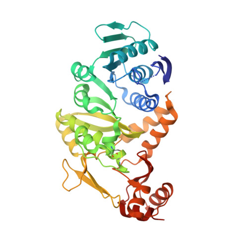
Crystal Structure of tRNA N(2),N(2)-Guanosine Dimethyltransferase Trm1 from Pyrococcus horikoshii

IhsanawatiNishimoto, M.Higashijima, K.Shirouzu, M.Grosjean, H.Bessho, Y.Yokoyama, S.

(2008) J Mol Biol 383: 871-884

  • DOI: https://doi.org/10.1016/j.jmb.2008.08.068
  • Primary Citation of Related Structures:  
    2DUL, 2EJT, 2EJU, 2YTZ

  • PubMed Abstract: 

    Trm1 catalyzes a two-step reaction, leading to mono- and dimethylation of guanosine at position 26 in most eukaryotic and archaeal tRNAs. We report the crystal structures of Trm1 from Pyrococcus horikoshii liganded with S-adenosyl-l-methionine or S-adenosyl-l-homocysteine. The protein comprises N-terminal and C-terminal domains with class I methyltransferase and novel folds, respectively. The methyl moiety of S-adenosyl-l-methionine points toward the invariant Phe27 and Phe140 within a narrow pocket, where the target G26 might flip in. Mutagenesis of Phe27 or Phe140 to alanine abolished the enzyme activity, indicating their role in methylating G26. Structural analyses revealed that the movements of Phe140 and the loop preceding Phe27 may be involved in dissociation of the monomethylated tRNA*Trm1 complex prior to the second methylation. Moreover, the catalytic residues Asp138, Pro139, and Phe140 are in a different motif from that in DNA 6-methyladenosine methyltransferases, suggesting a different methyl transfer mechanism in the Trm1 family.


  • Organizational Affiliation

    Systems and Structural Biology Center, Yokohama Institute, RIKEN, 1-7-22 Suehiro, Tsurumi, Yokohama 230-0045, Japan.


Macromolecules
Find similar proteins by:  (by identity cutoff)  |  3D Structure
Entity ID: 1
MoleculeChains Sequence LengthOrganismDetailsImage
N(2),N(2)-dimethylguanosine tRNA methyltransferase378Pyrococcus horikoshiiMutation(s): 0 
Gene Names: trm1
EC: 2.1.1.32
UniProt
Find proteins for O59493 (Pyrococcus horikoshii (strain ATCC 700860 / DSM 12428 / JCM 9974 / NBRC 100139 / OT-3))
Explore O59493 
Go to UniProtKB:  O59493
Entity Groups  
Sequence Clusters30% Identity50% Identity70% Identity90% Identity95% Identity100% Identity
UniProt GroupO59493
Sequence Annotations
Expand
  • Reference Sequence
Small Molecules
Ligands 2 Unique
IDChains Name / Formula / InChI Key2D Diagram3D Interactions
SAH
Query on SAH

Download Ideal Coordinates CCD File 
B [auth A]S-ADENOSYL-L-HOMOCYSTEINE
C14 H20 N6 O5 S
ZJUKTBDSGOFHSH-WFMPWKQPSA-N
GOL
Query on GOL

Download Ideal Coordinates CCD File 
C [auth A]GLYCEROL
C3 H8 O3
PEDCQBHIVMGVHV-UHFFFAOYSA-N
Experimental Data & Validation

Experimental Data

  • Method: X-RAY DIFFRACTION
  • Resolution: 1.95 Å
  • R-Value Free: 0.246 
  • R-Value Work: 0.200 
  • R-Value Observed: 0.200 
  • Space Group: C 2 2 21
Unit Cell:
Length ( Å )Angle ( ˚ )
a = 74.781α = 90
b = 109.655β = 90
c = 96.51γ = 90
Software Package:
Software NamePurpose
CNSrefinement
HKL-2000data collection
HKL-2000data reduction
HKL-2000data scaling
MOLREPphasing

Structure Validation

View Full Validation Report


Ligand Structure Quality Assessment 


Entry History 

Deposition Data

Revision History  (Full details and data files)

  • Version 1.0: 2007-09-25
    Type: Initial release
  • Version 1.1: 2011-07-13
    Changes: Non-polymer description, Version format compliance
  • Version 1.2: 2023-10-25
    Changes: Data collection, Database references, Derived calculations, Refinement description