2E3X | pdb_00002e3x

Crystal structure of Russell's viper venom metalloproteinase


Experimental Data Snapshot

  • Method: X-RAY DIFFRACTION
  • Resolution: 2.91 Å
  • R-Value Free: 
    0.273 (Depositor), 0.270 (DCC) 
  • R-Value Work: 
    0.218 (Depositor), 0.222 (DCC) 

Starting Models: experimental
View more details

wwPDB Validation 3D Report Full Report

Validation slider image for 2E3X

Ligand Structure Quality Assessment 


This is version 3.2 of the entry. See complete history

Literature

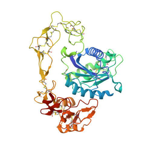
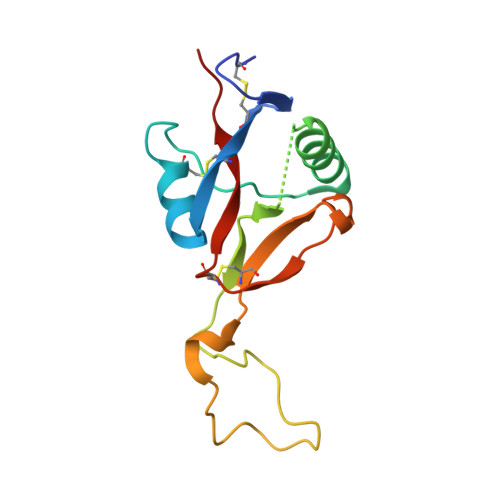
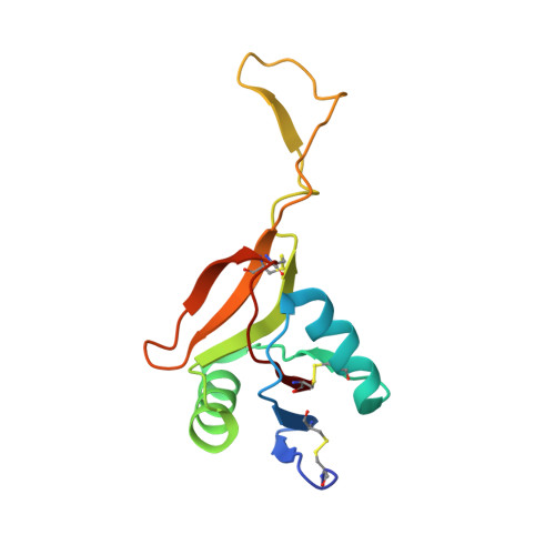
Crystal structure of RVV-X: an example of evolutionary gain of specificity by ADAM proteinases.

Takeda, S.Igarashi, T.Mori, H.

(2007) FEBS Lett 581: 5859-5864

  • DOI: https://doi.org/10.1016/j.febslet.2007.11.062
  • Primary Citation Related Structures: 
    2E3X

  • PubMed Abstract: 

    Russell's viper venom factor X activator (RVV-X) is a heterotrimeric metalloproteinase with a mammalian ADAM-like heavy chain and two lectin-like light chains. The crystal structure of RVV-X has been determined at 2.9 A resolution and shows a hook-spanner-wrench-like architecture, in which the metalloproteinase/disintegrin region constitutes a hook, and the lectin-like domains constitute a handle. A 6.5nm separation between the catalytic site and a putative exosite suggests a docking model for factor X. The structure provides a typical example of the molecular evolution of multi-subunit proteins and insights into the molecular basis of target recognition and proteolysis by ADAM/adamalysin/reprolysin proteinases.


  • Organizational Affiliation
    • Department of Cardiac Physiology, National Cardiovascular Center Research Institute, 5-7-1 Fujishiro-dai, Suita, Osaka 565-8565, Japan. stakeda@ri.ncvc.go.jp

Macromolecule Content 

  • Total Structure Weight: 80.29 kDa 
  • Atom Count: 5,438 
  • Modeled Residue Count: 665 
  • Deposited Residue Count: 683 
  • Unique protein chains: 3

Macromolecules

Find similar proteins by:|  3D Structure
Entity ID: 1
MoleculeChains  Sequence LengthOrganismDetailsImage
Coagulation factor X-activating enzyme heavy chain427Daboia siamensisMutation(s): 0 
EC: 3.4.24.58
UniProt
Find proteins for Q7LZ61 (Daboia siamensis)
Explore Q7LZ61 
Go to UniProtKB:  Q7LZ61
Entity Groups
Sequence Clusters30% Identity50% Identity70% Identity90% Identity95% Identity100% Identity
UniProt GroupQ7LZ61
Glycosylation
Glycosylation Sites: 2
Sequence Annotations
Expand
Reference Sequence
Find similar proteins by:|  3D Structure
Entity ID: 2
MoleculeChains  Sequence LengthOrganismDetailsImage
Coagulation factor X-activating enzyme light chain 2134Daboia siamensisMutation(s): 0 
UniProt
Find proteins for Q4PRD2 (Daboia siamensis)
Explore Q4PRD2 
Go to UniProtKB:  Q4PRD2
Entity Groups
Sequence Clusters30% Identity50% Identity70% Identity90% Identity95% Identity100% Identity
UniProt GroupQ4PRD2
Glycosylation
Glycosylation Sites: 1
Sequence Annotations
Expand
Reference Sequence
Find similar proteins by:|  3D Structure
Entity ID: 3
MoleculeChains  Sequence LengthOrganismDetailsImage
Coagulation factor X-activating enzyme light chain 1122Daboia siamensisMutation(s): 0 
UniProt
Find proteins for Q4PRD1 (Daboia siamensis)
Explore Q4PRD1 
Go to UniProtKB:  Q4PRD1
Entity Groups
Sequence Clusters30% Identity50% Identity70% Identity90% Identity95% Identity100% Identity
UniProt GroupQ4PRD1
Glycosylation
Glycosylation Sites: 1
Sequence Annotations
Expand
Reference Sequence

Oligosaccharides

Help  
Entity ID: 4
MoleculeChains Length2D Diagram GlycosylationD Interactions
2-acetamido-2-deoxy-beta-D-glucopyranose-(1-4)-2-acetamido-2-deoxy-beta-D-glucopyranose
D
2N-Glycosylation
Glycosylation Resources
GlyTouCan: G42666HT
GlyCosmos: G42666HT
GlyGen: G42666HT
Entity ID: 5
MoleculeChains Length2D Diagram GlycosylationD Interactions
alpha-D-mannopyranose-(1-6)-alpha-D-mannopyranose-(1-4)-2-acetamido-2-deoxy-beta-D-glucopyranose-(1-4)-2-acetamido-2-deoxy-beta-D-glucopyranose
E
4N-Glycosylation
Glycosylation Resources
GlyTouCan: G04854NQ
GlyCosmos: G04854NQ
GlyGen: G04854NQ

Small Molecules

Ligands 4 Unique
IDChains Name / Formula / InChI Key2D Diagram3D Interactions
GM6

Query on GM6


Download:Ideal Coordinates CCD File
L [auth A]3-(N-HYDROXYCARBOXAMIDO)-2-ISOBUTYLPROPANOYL-TRP-METHYLAMIDE
C20 H28 N4 O4
NITYDPDXAAFEIT-DYVFJYSZSA-N
NAG

Query on NAG


Download:Ideal Coordinates CCD File
F [auth A],
M [auth B]
2-acetamido-2-deoxy-beta-D-glucopyranose
C8 H15 N O6
OVRNDRQMDRJTHS-FMDGEEDCSA-N
ZN

Query on ZN


Download:Ideal Coordinates CCD File
G [auth A]ZINC ION
Zn
PTFCDOFLOPIGGS-UHFFFAOYSA-N
CA

Query on CA


Download:Ideal Coordinates CCD File
H [auth A],
I [auth A],
J [auth A],
K [auth A],
N [auth B]
CALCIUM ION
Ca
BHPQYMZQTOCNFJ-UHFFFAOYSA-N

Experimental Data & Validation

Experimental Data

  • Method: X-RAY DIFFRACTION
  • Resolution: 2.91 Å
  • R-Value Free:  0.273 (Depositor), 0.270 (DCC) 
  • R-Value Work:  0.218 (Depositor), 0.222 (DCC) 
Space Group: P 21 21 21
Unit Cell:
Length ( Å )Angle ( ˚ )
a = 70.35α = 90
b = 91.73β = 90
c = 152.93γ = 90
Software Package:
Software NamePurpose
ADSCdata collection
MOLREPphasing
CNSrefinement
HKL-2000data reduction
HKL-2000data scaling

Structure Validation

View Full Validation Report


Ligand Structure Quality Assessment 


Entry History 

Deposition Data

Revision History  (Full details and data files)

  • Version 1.0: 2007-12-11
    Type: Initial release
  • Version 1.1: 2011-07-13
    Changes: Derived calculations, Version format compliance
  • Version 2.0: 2019-12-25
    Changes: Advisory, Atomic model, Data collection, Database references, Derived calculations
  • Version 3.0: 2020-07-29
    Type: Remediation
    Reason: Carbohydrate remediation
    Changes: Advisory, Atomic model, Data collection, Derived calculations, Structure summary
  • Version 3.1: 2023-10-25
    Changes: Data collection, Database references, Derived calculations, Refinement description, Structure summary
  • Version 3.2: 2024-10-23
    Changes: Structure summary