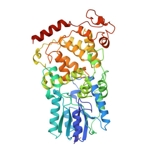
2E0I

Crystal structure of archaeal photolyase from Sulfolobus tokodaii with two FAD molecules: Implication of a novel light-harvesting cofactor


Experimental Data Snapshot

  • Method: X-RAY DIFFRACTION
  • Resolution: 2.80 Å
  • R-Value Free: 0.247 
  • R-Value Work: 0.223 

wwPDB Validation   3D Report Full Report


This is version 1.3 of the entry. See complete history


Literature

Crystal Structure of Archaeal Photolyase from Sulfolobus tokodaii with Two FAD Molecules: Implication of a Novel Light-harvesting Cofactor

Fujihashi, M.Numoto, N.Kobayashi, Y.Mizushima, A.Tsujimura, M.Nakamura, A.Kawarabayasi, Y.Miki, K.

(2007) J Mol Biol 365: 903-910

  • DOI: https://doi.org/10.1016/j.jmb.2006.10.012
  • Primary Citation of Related Structures:  
    2E0I

  • PubMed Abstract: 

    UV exposure of DNA molecules induces serious DNA lesions. The cyclobutane pyrimidine dimer (CPD) photolyase repairs CPD-type - lesions by using the energy of visible light. Two chromophores for different roles have been found in this enzyme family; one catalyzes the CPD repair reaction and the other works as an antenna pigment that harvests photon energy. The catalytic cofactor of all known photolyases is FAD, whereas several light-harvesting cofactors are found. Currently, 5,10-methenyltetrahydrofolate (MTHF), 8-hydroxy-5-deaza-riboflavin (8-HDF) and FMN are the known light-harvesting cofactors, and some photolyases lack the chromophore. Three crystal structures of photolyases from Escherichia coli (Ec-photolyase), Anacystis nidulans (An-photolyase), and Thermus thermophilus (Tt-photolyase) have been determined; however, no archaeal photolyase structure is available. A similarity search of archaeal genomic data indicated the presence of a homologous gene, ST0889, on Sulfolobus tokodaii strain7. An enzymatic assay reveals that ST0889 encodes photolyase from S. tokodaii (St-photolyase). We have determined the crystal structure of the St-photolyase protein to confirm its structural features and to investigate the mechanism of the archaeal DNA repair system with light energy. The crystal structure of the St-photolyase is superimposed very well on the three known photolyases including the catalytic cofactor FAD. Surprisingly, another FAD molecule is found at the position of the light-harvesting cofactor. This second FAD molecule is well accommodated in the crystal structure, suggesting that FAD works as a novel light-harvesting cofactor of photolyase. In addition, two of the four CPD recognition residues in the crystal structure of An-photolyase are not found in St-photolyase, which might utilize a different mechanism to recognize the CPD from that of An-photolyase.


  • Organizational Affiliation

    Department of Chemistry, Graduate School of Science, Kyoto University, Sakyo-ku, Kyoto 606-8502, Japan.


Macromolecules
Find similar proteins by:  (by identity cutoff)  |  3D Structure
Entity ID: 1
MoleculeChains Sequence LengthOrganismDetailsImage
432aa long hypothetical deoxyribodipyrimidine photolyase
A, B, C, D
440Sulfurisphaera tokodaii str. 7Mutation(s): 0 
Gene Names: ST0889
EC: 4.1.99.3
UniProt
Find proteins for F9VNB1 (Sulfurisphaera tokodaii (strain DSM 16993 / JCM 10545 / NBRC 100140 / 7))
Explore F9VNB1 
Go to UniProtKB:  F9VNB1
Entity Groups  
Sequence Clusters30% Identity50% Identity70% Identity90% Identity95% Identity100% Identity
UniProt GroupF9VNB1
Sequence Annotations
Expand
  • Reference Sequence
Small Molecules
Ligands 2 Unique
IDChains Name / Formula / InChI Key2D Diagram3D Interactions
FAD
Query on FAD

Download Ideal Coordinates CCD File 
E [auth A]
F [auth A]
I [auth B]
J [auth B]
L [auth C]
E [auth A],
F [auth A],
I [auth B],
J [auth B],
L [auth C],
M [auth C],
P [auth D],
Q [auth D]
FLAVIN-ADENINE DINUCLEOTIDE
C27 H33 N9 O15 P2
VWWQXMAJTJZDQX-UYBVJOGSSA-N
MPD
Query on MPD

Download Ideal Coordinates CCD File 
G [auth A]
H [auth A]
K [auth B]
N [auth C]
O [auth C]
G [auth A],
H [auth A],
K [auth B],
N [auth C],
O [auth C],
R [auth D]
(4S)-2-METHYL-2,4-PENTANEDIOL
C6 H14 O2
SVTBMSDMJJWYQN-YFKPBYRVSA-N
Experimental Data & Validation

Experimental Data

  • Method: X-RAY DIFFRACTION
  • Resolution: 2.80 Å
  • R-Value Free: 0.247 
  • R-Value Work: 0.223 
  • Space Group: P 1 21 1
Unit Cell:
Length ( Å )Angle ( ˚ )
a = 79.875α = 90
b = 114.715β = 91.49
c = 167.684γ = 90
Software Package:
Software NamePurpose
CNSrefinement
HKL-2000data collection
HKL-2000data reduction
SCALEPACKdata scaling
MOLREPphasing

Structure Validation

View Full Validation Report


Entry History 

Deposition Data

Revision History  (Full details and data files)

  • Version 1.0: 2006-11-28
    Type: Initial release
  • Version 1.1: 2008-04-30
    Changes: Version format compliance
  • Version 1.2: 2011-07-13
    Changes: Source and taxonomy, Version format compliance
  • Version 1.3: 2023-10-25
    Changes: Data collection, Database references, Derived calculations, Refinement description