


Crystal structure of MS0666
Wang, H., Kishishita, S., Murayama, K., Terada, T., Shirouzu, M., Yokoyama, S.To be published.
Experimental Data Snapshot
wwPDB Validation 3D Report Full Report

Entity ID: 1 | |||||
|---|---|---|---|---|---|
| Molecule | Chains | Sequence Length | Organism | Details | Image |
| RAL GUANINE NUCLEOTIDE EXCHANGE FACTOR RALGPS1A | 126 | Mus musculus | Mutation(s): 3 Membrane Entity: Yes | 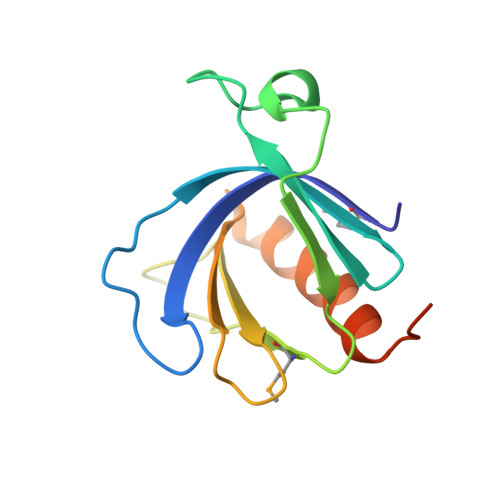 | |
UniProt & NIH Common Fund Data Resources | |||||
IMPC: MGI:1922008 | |||||
Entity Groups | |||||
| Sequence Clusters | 30% Identity50% Identity70% Identity90% Identity95% Identity100% Identity | ||||
| UniProt Group | A2AR50 | ||||
Sequence AnnotationsExpand | |||||
Reference Sequence | |||||
| Modified Residues 1 Unique | |||||
|---|---|---|---|---|---|
| ID | Chains | Type | Formula | 2D Diagram | Parent |
| MSE Query on MSE | A, B | L-PEPTIDE LINKING | C5 H11 N O2 Se |  | MET |
| Length ( Å ) | Angle ( ˚ ) |
|---|---|
| a = 43.68 | α = 90 |
| b = 61.077 | β = 104.72 |
| c = 43.939 | γ = 90 |
| Software Name | Purpose |
|---|---|
| CNS | refinement |
| HKL-2000 | data reduction |
| HKL-2000 | data scaling |
| SOLVE | phasing |


