2DPI

Ternary complex of hPoli with DNA and dCTP


Experimental Data Snapshot

  • Method: X-RAY DIFFRACTION
  • Resolution: 2.30 Å
  • R-Value Free: 0.278 
  • R-Value Work: 0.228 
  • R-Value Observed: 0.232 

wwPDB Validation   3D Report Full Report


Ligand Structure Quality Assessment 


This is version 1.3 of the entry. See complete history


Literature

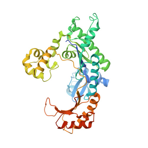
Hoogsteen base pair formation promotes synthesis opposite the 1,N(6)-ethenodeoxyadenosine lesion by human DNA polymerase iota.

Nair, D.T.Johnson, R.E.Prakash, L.Prakash, S.Aggarwal, A.K.

(2006) Nat Struct Mol Biol 13: 619-625

  • DOI: https://doi.org/10.1038/nsmb1118
  • Primary Citation of Related Structures:  
    2DPI, 2DPJ

  • PubMed Abstract: 

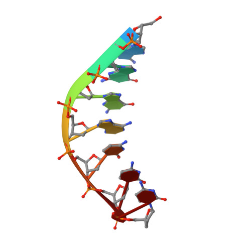
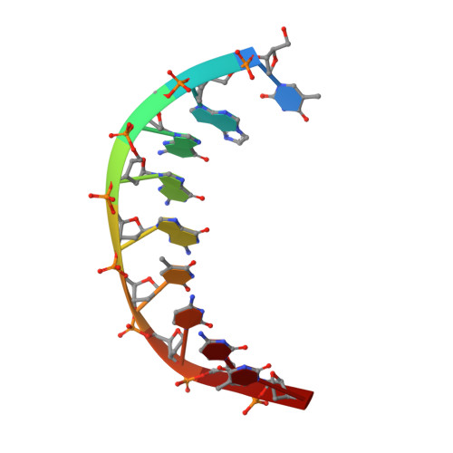
    The 1,N6-ethenodeoxyadenosine (epsilon dA) lesion is promutagenic and has been implicated in carcinogenesis. We show here that human Pol iota, a Y-family DNA polymerase, can promote replication through this lesion by proficiently incorporating a nucleotide opposite it. The structural basis of this action is rotation of the epsilon dA adduct to the syn conformation in the Pol iota active site and presentation of its 'Hoogsteen edge' for hydrogen-bonding with incoming dTTP or dCTP. We also show that Pol zeta carries out the subsequent extension reaction and that efficiency of extension from epsilon dA x T is notably higher than from epsilon dA x C. Together, our studies reveal for the first time how the exocyclic epsilon dA adduct is accommodated in a DNA polymerase active site, and they show that the combined action of Pol iota and Pol zeta provides for efficient and error-free synthesis through this potentially carcinogenic DNA lesion.


  • Organizational Affiliation

    Department of Molecular Physiology and Biophysics, Mount Sinai School of Medicine, Box 1677, 1425 Madison Avenue, New York, New York 10029, USA.


Macromolecules

Find similar proteins by:  (by identity cutoff)  |  3D Structure
Entity ID: 3
MoleculeChains Sequence LengthOrganismDetailsImage
DNA polymerase iotaC [auth A]420Homo sapiensMutation(s): 0 
Gene Names: RAD30 homolog BEta2
EC: 2.7.7.7
UniProt & NIH Common Fund Data Resources
Find proteins for Q9UNA4 (Homo sapiens)
Explore Q9UNA4 
Go to UniProtKB:  Q9UNA4
PHAROS:  Q9UNA4
GTEx:  ENSG00000101751 
Entity Groups  
Sequence Clusters30% Identity50% Identity70% Identity90% Identity95% Identity100% Identity
UniProt GroupQ9UNA4
Sequence Annotations
Expand
  • Reference Sequence

Find similar nucleic acids by:  Sequence   |   3D Structure  

Entity ID: 1
MoleculeChains LengthOrganismImage
5'-D(*AP*GP*GP*AP*CP*CP*(DOC))-3'A [auth P]7N/A
Sequence Annotations
Expand
  • Reference Sequence

Find similar nucleic acids by:  Sequence   |   3D Structure  

Entity ID: 2
MoleculeChains LengthOrganismImage
5'-D(*TP*(EDA)P*GP*GP*GP*TP*CP*CP*T)-3'B [auth T]9N/A
Sequence Annotations
Expand
  • Reference Sequence
Experimental Data & Validation

Experimental Data

  • Method: X-RAY DIFFRACTION
  • Resolution: 2.30 Å
  • R-Value Free: 0.278 
  • R-Value Work: 0.228 
  • R-Value Observed: 0.232 
  • Space Group: P 65 2 2
Unit Cell:
Length ( Å )Angle ( ˚ )
a = 98.148α = 90
b = 98.148β = 90
c = 203.112γ = 120
Software Package:
Software NamePurpose
REFMACrefinement
DENZOdata reduction
SCALEPACKdata scaling
CNSphasing

Structure Validation

View Full Validation Report


Ligand Structure Quality Assessment 


Entry History 

Deposition Data

Revision History  (Full details and data files)

  • Version 1.0: 2006-07-04
    Type: Initial release
  • Version 1.1: 2008-04-30
    Changes: Version format compliance
  • Version 1.2: 2011-07-13
    Changes: Advisory, Version format compliance
  • Version 1.3: 2023-10-25
    Changes: Data collection, Database references, Derived calculations, Refinement description