2DOR

DIHYDROOROTATE DEHYDROGENASE A FROM LACTOCOCCUS LACTIS COMPLEXED WITH OROTATE


Experimental Data Snapshot

  • Method: X-RAY DIFFRACTION
  • Resolution: 2.00 Å
  • R-Value Free: 0.187 
  • R-Value Work: 0.161 
  • R-Value Observed: 0.161 

wwPDB Validation   3D Report Full Report


Ligand Structure Quality Assessment 


This is version 1.2 of the entry. See complete history


Literature

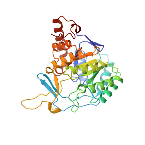
The crystal structure of Lactococcus lactis dihydroorotate dehydrogenase A complexed with the enzyme reaction product throws light on its enzymatic function.

Rowland, P.Bjornberg, O.Nielsen, F.S.Jensen, K.F.Larsen, S.

(1998) Protein Sci 7: 1269-1279

  • DOI: https://doi.org/10.1002/pro.5560070601
  • Primary Citation of Related Structures:  
    2DOR

  • PubMed Abstract: 
  • Dihydroorotate dehydrogenases (DHODs) catalyze the oxidation of (S)-dihydroorotate to orotate, the fourth step and only redox reaction in the de novo biosynthesis of pyrimidine nucleotides. A description is given of the crystal structure of Lactococcus lactis dihydroorotate dehydrogenase A (DHODA) complexed with the product of the enzyme reaction orotate ...

    Dihydroorotate dehydrogenases (DHODs) catalyze the oxidation of (S)-dihydroorotate to orotate, the fourth step and only redox reaction in the de novo biosynthesis of pyrimidine nucleotides. A description is given of the crystal structure of Lactococcus lactis dihydroorotate dehydrogenase A (DHODA) complexed with the product of the enzyme reaction orotate. The structure of the complex to 2.0 A resolution has been compared with the structure of the native enzyme. The active site of DHODA is known to contain a water filled cavity buried beneath a highly conserved and flexible loop. In the complex the orotate displaces the water molecules from the active site and stacks above the DHODA flavin isoalloxazine ring, causing only small movements of the surrounding protein residues. The orotate is completely buried beneath the protein surface, and the orotate binding causes a significant reduction in the mobility of the active site loop. The orotate is bound by four conserved asparagine side chains (Asn 67, Asn 127, Asn 132, and Asn 193), the side chains of Lys 43 and Ser 194, and the main chain NH groups of Met 69, Gly 70, and Leu 71. Of these the Lys 43 side chain makes hydrogen bonds to both the flavin isoalloxazine ring and the carboxylate group of the orotate. Potential interactions with bound dihydroorotate are considered using the orotate complex as a basis for molecular modeling. The role of Cys 130 as the active site base is discussed, and the sequence conservation of the active site residues across the different families of DHODs is reviewed, along with implications for differences in substrate binding and in the catalytic mechanisms between these families.


    Organizational Affiliation

    Centre for Crystallographic Studies, Department of Chemistry, University of Copenhagen, Denmark.


Macromolecules
Find similar proteins by:  (by identity cutoff)  |  3D Structure
Entity ID: 1
MoleculeChainsSequence LengthOrganismDetailsImage
DIHYDROOROTATE DEHYDROGENASE AA, B311Lactococcus lactisMutation(s): 0 
EC: 1.3.3.1 (PDB Primary Data), 1.3.98.1 (UniProt)
UniProt
Find proteins for A2RJT9 (Lactococcus lactis subsp. cremoris (strain MG1363))
Explore A2RJT9 
Go to UniProtKB:  A2RJT9
Entity Groups  
Sequence Clusters30% Identity50% Identity70% Identity90% Identity95% Identity100% Identity
UniProt GroupA2RJT9
Protein Feature View
Expand
  • Reference Sequence
Small Molecules
Ligands 2 Unique
IDChainsName / Formula / InChI Key2D Diagram3D Interactions
FMN
Query on FMN

Download Ideal Coordinates CCD File 
C [auth A],
E [auth B]
FLAVIN MONONUCLEOTIDE
C17 H21 N4 O9 P
FVTCRASFADXXNN-SCRDCRAPSA-N
 Ligand Interaction
ORO
Query on ORO

Download Ideal Coordinates CCD File 
D [auth A],
F [auth B]
OROTIC ACID
C5 H4 N2 O4
PXQPEWDEAKTCGB-UHFFFAOYSA-N
 Ligand Interaction
Experimental Data & Validation

Experimental Data

  • Method: X-RAY DIFFRACTION
  • Resolution: 2.00 Å
  • R-Value Free: 0.187 
  • R-Value Work: 0.161 
  • R-Value Observed: 0.161 
  • Space Group: P 1 21 1
Unit Cell:
Length ( Å )Angle ( ˚ )
a = 53.91α = 90
b = 109.03β = 104.43
c = 67.07γ = 90
Software Package:
Software NamePurpose
X-PLORmodel building
X-PLORrefinement
DENZOdata reduction
SCALEPACKdata scaling
X-PLORphasing

Structure Validation

View Full Validation Report


Ligand Structure Quality Assessment 


Entry History 

Deposition Data

Revision History  (Full details and data files)

  • Version 1.0: 1998-08-12
    Type: Initial release
  • Version 1.1: 2008-03-24
    Changes: Version format compliance
  • Version 1.2: 2011-07-13
    Changes: Version format compliance