2DM5 | pdb_00002dm5

Thermodynamic Penalty Arising From Burial of a Ligand Polar Group Within a Hydrophobic Pocket of a Protein Receptor


Experimental Data Snapshot

  • Method: X-RAY DIFFRACTION
  • Resolution: 1.70 Å
  • R-Value Free: 
    0.200 (Depositor), 0.190 (DCC) 
  • R-Value Work: 
    0.190 (Depositor), 0.191 (DCC) 
  • R-Value Observed: 
    0.190 (Depositor) 

Starting Model: experimental
View more details

wwPDB Validation 3D Report Full Report

Validation slider image for 2DM5

This is version 1.4 of the entry. See complete history

Literature

Thermodynamic penalty arising from burial of a ligand polar group within a hydrophobic pocket of a protein receptor

Barratt, E.Bronowska, A.Vondrasek, J.Cerny, J.Bingham, R.Phillips, S.Homans, S.W.

(2006) J Mol Biology 362: 994-1003

  • DOI: https://doi.org/10.1016/j.jmb.2006.07.067
  • Primary Citation Related Structures: 
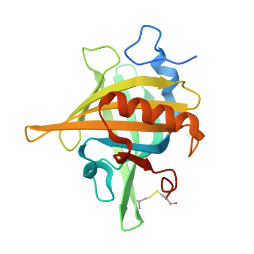
    2DM5

  • PubMed Abstract: 

    Here, we examine the thermodynamic penalty arising from burial of a polar group in a hydrophobic pocket that forms part of the binding-site of the major urinary protein (MUP-I). X-ray crystal structures of the complexes of octanol, nonanol and 1,8 octan-diol indicate that these ligands bind with similar orientations in the binding pocket. Each complex is characterised by a bridging water molecule between the hydroxyl group of Tyr120 and the hydroxyl group of each ligand. The additional hydroxyl group of 1,8 octan-diol is thereby forced to reside in a hydrophobic pocket, and isothermal titration calorimetry experiments indicate that this is accompanied by a standard free energy penalty of +21 kJ/mol with respect to octanol and +18 kJ/mol with respect to nonanol. Consideration of the solvation thermodynamics of each ligand enables the "intrinsic" (solute-solute) interaction energy to be determined, which indicates a favourable enthalpic component and an entropic component that is small or zero. These data indicate that the thermodynamic penalty to binding derived from the unfavourable desolvation of 1,8 octan-diol is partially offset by a favourable intrinsic contribution. Quantum chemical calculations suggest that this latter contribution derives from favourable solute-solute dispersion interactions.


  • Organizational Affiliation
    • Astbury Centre for Structural Molecular Biology, School of Biochemistry and Molecular Biology, University of Leeds, LS2 9JT, UK.

Macromolecule Content 

  • Total Structure Weight: 20.74 kDa 
  • Atom Count: 1,460 
  • Modeled Residue Count: 157 
  • Deposited Residue Count: 174 
  • Unique protein chains: 1

Macromolecules

Find similar proteins by:|  3D Structure
Entity ID: 1
MoleculeChains  Sequence LengthOrganismDetailsImage
Major Urinary Protein174Mus musculusMutation(s): 0 
Gene Names: MUP1
UniProt
Find proteins for P11588 (Mus musculus)
Explore P11588 
Go to UniProtKB:  P11588
Entity Groups
Sequence Clusters30% Identity50% Identity70% Identity90% Identity95% Identity100% Identity
UniProt GroupP11588
Sequence Annotations
Expand
Reference Sequence

Experimental Data & Validation

Experimental Data

  • Method: X-RAY DIFFRACTION
  • Resolution: 1.70 Å
  • R-Value Free:  0.200 (Depositor), 0.190 (DCC) 
  • R-Value Work:  0.190 (Depositor), 0.191 (DCC) 
  • R-Value Observed: 0.190 (Depositor) 
Space Group: P 43 21 2
Unit Cell:
Length ( Å )Angle ( ˚ )
a = 53.522α = 90
b = 53.522β = 90
c = 137.192γ = 90
Software Package:
Software NamePurpose
CrystalCleardata collection
SCALAdata scaling
CNSrefinement
CrystalCleardata reduction
CCP4data scaling
CNSphasing

Structure Validation

View Full Validation Report


Entry History 

Deposition Data

Revision History  (Full details and data files)

  • Version 1.0: 2006-10-17
    Type: Initial release
  • Version 1.1: 2008-04-30
    Changes: Version format compliance
  • Version 1.2: 2011-07-13
    Changes: Version format compliance
  • Version 1.3: 2023-10-25
    Changes: Data collection, Database references, Derived calculations, Refinement description
  • Version 1.4: 2024-10-23
    Changes: Structure summary