2DKN

Crystal structure of the 3-alpha-hydroxysteroid dehydrogenase from Pseudomonas sp. B-0831 complexed with NADH


Experimental Data Snapshot

  • Method: X-RAY DIFFRACTION
  • Resolution: 1.80 Å
  • R-Value Free: 0.205 
  • R-Value Work: 0.172 
  • R-Value Observed: 0.174 

wwPDB Validation   3D Report Full Report


Ligand Structure Quality Assessment 


This is version 1.3 of the entry. See complete history


Literature

Apo- and Holo-structures of 3{alpha}-Hydroxysteroid Dehydrogenase from Pseudomonas sp. B-0831: LOOP-HELIX TRANSITION INDUCED BY COENZYME BINDING

Nakamura, S.Oda, M.Kataoka, S.Ueda, S.Uchiyama, S.Yoshida, T.Kobayashi, Y.Ohkubo, T.

(2006) J Biol Chem 281: 31876-31884

  • DOI: https://doi.org/10.1074/jbc.M604226200
  • Primary Citation of Related Structures:  
    2DKN

  • PubMed Abstract: 

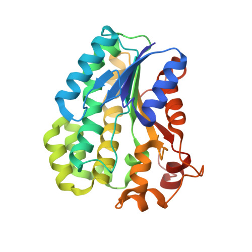
    Bacterial 3alpha-hydroxysteroid dehydrogenase, which belongs to a short-chain dehydrogenase/reductase family and forms a dimer composed of two 26-kDa subunits, catalyzes the oxidoreduction of hydroxysteroids in a coenzyme-dependent manner. This enzyme also catalyzes the oxidoreduction of nonsteroid compounds that play an important role in xenobiotic metabolism of bacteria. We performed an x-ray analysis on the crystal of Ps3alphaHSD, the enzyme from Pseudomonas sp. B-0831 complexed with NADH. The resulting crystal structure at 1.8A resolution showed that Ps3alphaHSD exists as a structural heterodimer composed of apo- and holo-subunits. A distinct structural difference between them was found in the 185-207-amino acid region, where the structure in the apo-subunit is disordered whereas that in the holo-subunit consists of two alpha-helices. This fact proved that the NADH binding allows the helical structures to form the substrate binding pocket even in the absence of the substrate, although the region corresponds to the so-called "substrate-binding loop." The induction of alpha-helices in solution by the coenzyme binding was also confirmed by the CD experiment. In addition, the CD experiment revealed that the helix-inducing ability of NADH is stronger than that of NAD. We discuss the negative cooperativity for the coenzyme binding, which is caused by the effect of the structural change transferred between the subunits of the heterodimer.


  • Organizational Affiliation

    Graduate School of Pharmaceutical Sciences, Osaka University, 1-6 Yamadaoka, Suita, Osaka 565-0871, Japan.


Macromolecules
Find similar proteins by:  (by identity cutoff)  |  3D Structure
Entity ID: 1
MoleculeChains Sequence LengthOrganismDetailsImage
3-alpha-hydroxysteroid dehydrogenase
A, B
255Pseudomonas sp. B-0831Mutation(s): 0 
EC: 1.1.1.50
UniProt
Find proteins for Q59718 (Pseudomonas sp. B-0831)
Explore Q59718 
Go to UniProtKB:  Q59718
Entity Groups  
Sequence Clusters30% Identity50% Identity70% Identity90% Identity95% Identity100% Identity
UniProt GroupQ59718
Sequence Annotations
Expand
  • Reference Sequence
Small Molecules
Ligands 1 Unique
IDChains Name / Formula / InChI Key2D Diagram3D Interactions
NAI
Query on NAI

Download Ideal Coordinates CCD File 
C [auth A]1,4-DIHYDRONICOTINAMIDE ADENINE DINUCLEOTIDE
C21 H29 N7 O14 P2
BOPGDPNILDQYTO-NNYOXOHSSA-N
Binding Affinity Annotations 
IDSourceBinding Affinity
NAI Binding MOAD:  2DKN Kd: 8800 (nM) from 1 assay(s)
Experimental Data & Validation

Experimental Data

  • Method: X-RAY DIFFRACTION
  • Resolution: 1.80 Å
  • R-Value Free: 0.205 
  • R-Value Work: 0.172 
  • R-Value Observed: 0.174 
  • Space Group: P 21 21 21
Unit Cell:
Length ( Å )Angle ( ˚ )
a = 62.457α = 90
b = 82.248β = 90
c = 86.569γ = 90
Software Package:
Software NamePurpose
REFMACrefinement
HKL-2000data reduction
HKL-2000data scaling
MOLREPphasing

Structure Validation

View Full Validation Report


Ligand Structure Quality Assessment 


Entry History 

Deposition Data

  • Released Date: 2006-08-15 
  • Deposition Author(s): Nakamura, S.

Revision History  (Full details and data files)

  • Version 1.0: 2006-08-15
    Type: Initial release
  • Version 1.1: 2008-04-30
    Changes: Version format compliance
  • Version 1.2: 2011-07-13
    Changes: Source and taxonomy, Version format compliance
  • Version 1.3: 2023-10-25
    Changes: Data collection, Database references, Derived calculations, Refinement description