2DHR

Whole cytosolic region of ATP-dependent metalloprotease FtsH (G399L)


Experimental Data Snapshot

  • Method: X-RAY DIFFRACTION
  • Resolution: 3.90 Å
  • R-Value Free: 0.342 
  • R-Value Work: 0.299 
  • R-Value Observed: 0.301 

wwPDB Validation   3D Report Full Report


Ligand Structure Quality Assessment 


This is version 1.3 of the entry. See complete history


Literature

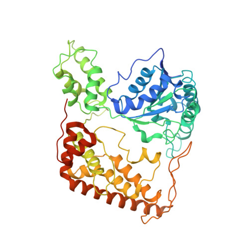
Structure of the Whole Cytosolic Region of ATP-Dependent Protease FtsH

Suno, R.Niwa, H.Tsuchiya, D.Zhang, X.Yoshida, M.Morikawa, K.

(2006) Mol Cell 22: 575-585

  • DOI: https://doi.org/10.1016/j.molcel.2006.04.020
  • Primary Citation of Related Structures:  
    2DHR, 2DI4, 4EIW

  • PubMed Abstract: 

    An ATP-dependent protease, FtsH, digests misassembled membrane proteins in order to maintain membrane integrity and digests short-lived soluble proteins in order to control their cellular regulation. This enzyme has an N-terminal transmembrane segment and a C-terminal cytosolic region consisting of an AAA+ ATPase domain and a protease domain. Here we present two crystal structures: the protease domain and the whole cytosolic region. The cytosolic region fully retains an ATP-dependent protease activity and adopts a three-fold-symmetric hexameric structure. The protease domains displayed a six-fold symmetry, while the AAA+ domains, each containing ADP, alternate two orientations relative to the protease domain, making "open" and "closed" interdomain contacts. Apparently, ATPase is active only in the closed form, and protease operates in the open form. The protease catalytic sites are accessible only through a tunnel following from the AAA+ domain of the adjacent subunit, raising a possibility of translocation of polypeptide substrate to the protease sites through this tunnel.


  • Organizational Affiliation

    Chemical Resources Laboratory, Tokyo Institute of Technology, 4259 Nagatsuta, Yokohama 226-8503, Japan.


Macromolecules
Find similar proteins by:  (by identity cutoff)  |  3D Structure
Entity ID: 1
MoleculeChains Sequence LengthOrganismDetailsImage
FtsH
A, B, C, D, E
A, B, C, D, E, F
499Thermus thermophilusMutation(s): 1 
UniProt
Find proteins for Q5SI82 (Thermus thermophilus (strain ATCC 27634 / DSM 579 / HB8))
Explore Q5SI82 
Go to UniProtKB:  Q5SI82
Entity Groups  
Sequence Clusters30% Identity50% Identity70% Identity90% Identity95% Identity100% Identity
UniProt GroupQ5SI82
Sequence Annotations
Expand
  • Reference Sequence
Experimental Data & Validation

Experimental Data

  • Method: X-RAY DIFFRACTION
  • Resolution: 3.90 Å
  • R-Value Free: 0.342 
  • R-Value Work: 0.299 
  • R-Value Observed: 0.301 
  • Space Group: P 31 2 1
Unit Cell:
Length ( Å )Angle ( ˚ )
a = 146.153α = 90
b = 146.153β = 90
c = 349.064γ = 120
Software Package:
Software NamePurpose
MOSFLMdata reduction
SCALAdata scaling
SHARPphasing
REFMACrefinement
CCP4data scaling

Structure Validation

View Full Validation Report


Ligand Structure Quality Assessment 


Entry History 

Deposition Data

Revision History  (Full details and data files)

  • Version 1.0: 2006-06-27
    Type: Initial release
  • Version 1.1: 2008-04-30
    Changes: Version format compliance
  • Version 1.2: 2011-07-13
    Changes: Version format compliance
  • Version 1.3: 2021-11-10
    Changes: Database references, Derived calculations