2D5R | pdb_00002d5r

Crystal Structure of a Tob-hCaf1 Complex


Experimental Data Snapshot

  • Method: X-RAY DIFFRACTION
  • Resolution: 2.50 Å
  • R-Value Free: 
    0.246 (Depositor) 
  • R-Value Work: 
    0.223 (Depositor) 
  • R-Value Observed: 
    0.225 (Depositor) 

wwPDB Validation 3D Report Full Report

Validation slider image for 2D5R

This is version 1.3 of the entry. See complete history

Literature

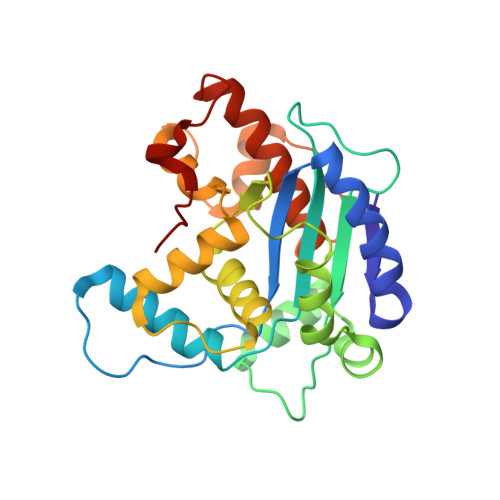
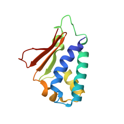
Structural basis for the antiproliferative activity of the Tob-hCaf1 complex.

Horiuchi, M.Takeuchi, K.Noda, N.Muroya, N.Suzuki, T.Nakamura, T.Kawamura-Tsuzuku, J.Takahasi, K.Yamamoto, T.Inagaki, F.

(2009) J Biological Chem 284: 13244-13255

  • DOI: https://doi.org/10.1074/jbc.M809250200
  • Primary Citation Related Structures: 
    2D5R

  • PubMed Abstract: 

    The Tob/BTG family is a group of antiproliferative proteins containing two highly homologous regions, Box A and Box B. These proteins all associate with CCR4-associated factor 1 (Caf1), which belongs to the ribonuclease D (RNase D) family of deadenylases and is a component of the CCR4-Not deadenylase complex. Here we determined the crystal structure of the complex of the N-terminal region of Tob and human Caf1 (hCaf1). Tob exhibited a novel fold, whereas hCaf1 most closely resembled the catalytic domain of yeast Pop2 and human poly(A)-specific ribonuclease. Interestingly, the association of hCaf1 was mediated by both Box A and Box B of Tob. Cell growth assays using both wild-type and mutant proteins revealed that deadenylase activity of Caf1 is not critical but complex formation is crucial to cell growth inhibition. Caf1 tethers Tob to the CCR4-Not deadenylase complex, and thereby Tob gathers several factors at its C-terminal region, such as poly(A)-binding proteins, to exert antiproliferative activity.


  • Organizational Affiliation
    • Department of Structural Biology, Graduate School of Pharmaceutical Sciences, Hokkaido University, Sapporo, Japan.

Macromolecule Content 

  • Total Structure Weight: 42.93 kDa 
  • Atom Count: 3,113 
  • Modeled Residue Count: 368 
  • Deposited Residue Count: 368 
  • Unique protein chains: 2

Macromolecules

Find similar proteins by:|  3D Structure
Entity ID: 1
MoleculeChains  Sequence LengthOrganismDetailsImage
CCR4-NOT transcription complex subunit 7252Homo sapiensMutation(s): 0 
EC: 3.1.13.4
UniProt & NIH Common Fund Data Resources
Find proteins for Q9UIV1 (Homo sapiens)
Explore Q9UIV1 
Go to UniProtKB:  Q9UIV1
PHAROS:  Q9UIV1
GTEx:  ENSG00000198791 
Entity Groups
Sequence Clusters30% Identity50% Identity70% Identity90% Identity95% Identity100% Identity
UniProt GroupQ9UIV1
Sequence Annotations
Expand
Reference Sequence
Find similar proteins by:|  3D Structure
Entity ID: 2
MoleculeChains  Sequence LengthOrganismDetailsImage
Tob1 protein116Homo sapiensMutation(s): 0 
UniProt & NIH Common Fund Data Resources
Find proteins for P50616 (Homo sapiens)
Explore P50616 
Go to UniProtKB:  P50616
PHAROS:  P50616
GTEx:  ENSG00000141232 
Entity Groups
Sequence Clusters30% Identity50% Identity70% Identity90% Identity95% Identity100% Identity
UniProt GroupP50616
Sequence Annotations
Expand
Reference Sequence

Experimental Data & Validation

Experimental Data

  • Method: X-RAY DIFFRACTION
  • Resolution: 2.50 Å
  • R-Value Free:  0.246 (Depositor) 
  • R-Value Work:  0.223 (Depositor) 
  • R-Value Observed: 0.225 (Depositor) 
Space Group: I 4 2 2
Unit Cell:
Length ( Å )Angle ( ˚ )
a = 151.505α = 90
b = 151.505β = 90
c = 114.181γ = 90
Software Package:
Software NamePurpose
CNSrefinement
MOSFLMdata reduction
CCP4data scaling
CNSphasing

Structure Validation

View Full Validation Report


Entry History 

Revision History  (Full details and data files)

  • Version 1.0: 2006-12-12
    Type: Initial release
  • Version 1.1: 2008-04-30
    Changes: Version format compliance
  • Version 1.2: 2011-07-13
    Changes: Version format compliance
  • Version 1.3: 2024-03-13
    Changes: Data collection, Database references