2D42

Crystal structure analysis of a non-toxic crystal protein from Bacillus thuringiensis


Experimental Data Snapshot

  • Method: X-RAY DIFFRACTION
  • Resolution: 2.07 Å
  • R-Value Free: 0.265 
  • R-Value Work: 0.230 
  • R-Value Observed: 0.230 

wwPDB Validation   3D Report Full Report


This is version 1.3 of the entry. See complete history


Literature

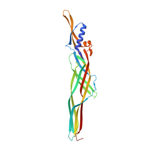
Nontoxic crystal protein from Bacillus thuringiensis demonstrates a remarkable structural similarity to beta-pore-forming toxins

Akiba, T.Higuchi, K.Mizuki, E.Ekino, K.Shin, T.Ohba, M.Kanai, R.Harata, K.

(2006) Proteins 63: 243-248


Macromolecules
Find similar proteins by:  (by identity cutoff)  |  3D Structure
Entity ID: 1
MoleculeChains Sequence LengthOrganismDetailsImage
non-toxic crystal protein
A, B
249Bacillus thuringiensisMutation(s): 0 
UniProt
Find proteins for Q6L5X8 (Bacillus thuringiensis serovar shandongiensis)
Explore Q6L5X8 
Go to UniProtKB:  Q6L5X8
Entity Groups  
Sequence Clusters30% Identity50% Identity70% Identity90% Identity95% Identity100% Identity
UniProt GroupQ6L5X8
Sequence Annotations
Expand
  • Reference Sequence
Experimental Data & Validation

Experimental Data

  • Method: X-RAY DIFFRACTION
  • Resolution: 2.07 Å
  • R-Value Free: 0.265 
  • R-Value Work: 0.230 
  • R-Value Observed: 0.230 
  • Space Group: P 31 1 2
Unit Cell:
Length ( Å )Angle ( ˚ )
a = 95.38α = 90
b = 95.38β = 90
c = 130.727γ = 120
Software Package:
Software NamePurpose
CNSrefinement
HKL-2000data reduction
SCALEPACKdata scaling
SHARPphasing

Structure Validation

View Full Validation Report


Entry History 

Deposition Data

Revision History  (Full details and data files)

  • Version 1.0: 2006-01-17
    Type: Initial release
  • Version 1.1: 2008-04-30
    Changes: Version format compliance
  • Version 1.2: 2011-07-13
    Changes: Version format compliance
  • Version 1.3: 2024-03-13
    Changes: Data collection, Database references