2D1G | pdb_00002d1g

Structure of Francisella tularensis Acid Phosphatase A (AcpA) bound to orthovanadate


Experimental Data Snapshot

  • Method: X-RAY DIFFRACTION
  • Resolution: 1.75 Å
  • R-Value Free: 
    0.231 (Depositor), 0.280 (DCC) 
  • R-Value Work: 
    0.198 (Depositor), 0.263 (DCC) 

wwPDB Validation 3D Report Full Report

Validation slider image for 2D1G

This is version 1.4 of the entry. See complete history

Literature

Structure of Francisella tularensis AcpA: prototype of a unique superfamily of acid phosphatases and phospholipases C

Felts, R.L.Reilly, T.J.Tanner, J.J.

(2006) J Biological Chem 281: 30289-30298

  • DOI: https://doi.org/10.1074/jbc.M606391200
  • Primary Citation Related Structures: 
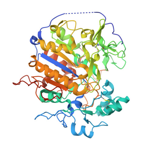
    2D1G

  • PubMed Abstract: 

    AcpA is a respiratory burst-inhibiting acid phosphatase from the Centers for Disease Control and Prevention Category A bioterrorism agent Francisella tularensis and prototype of a superfamily of acid phosphatases and phospholipases C. We report the 1.75-A resolution crystal structure of AcpA complexed with the inhibitor orthovanadate, which is the first structure of any F. tularensis protein and the first for any member of this superfamily. The core domain is a twisted 8-stranded beta-sheet flanked by three alpha-helices on either side, with the active site located above the carboxyl-terminal edge of the beta-sheet. This architecture is unique among acid phosphatases and resembles that of alkaline phosphatase. Unexpectedly, the active site features a serine nucleophile and metal ion with octahedral coordination. Structure-based sequence analysis of the AcpA superfamily predicts that the hydroxyl nucleophile and metal center are also present in AcpA-like phospholipases C. These results imply a phospholipase C catalytic mechanism that is radically different from that of zinc metallophospholipases.


  • Organizational Affiliation
    • Department of Chemistry, University of Missouri, Columbia, Missouri 65211, USA.

Macromolecule Content 

  • Total Structure Weight: 114.46 kDa 
  • Atom Count: 8,182 
  • Modeled Residue Count: 953 
  • Deposited Residue Count: 996 
  • Unique protein chains: 1

Macromolecules

Find similar proteins by:|  3D Structure
Entity ID: 1
MoleculeChains  Sequence LengthOrganismDetailsImage
acid phosphatase
A, B
498Francisella tularensis subsp. novicidaMutation(s): 0 
Gene Names: acpA
EC: 3.1.3.2
UniProt
Find proteins for A0Q436 (Francisella tularensis subsp. novicida (strain ATCC 15482 / CCUG 33449 / U112))
Explore A0Q436 
Go to UniProtKB:  A0Q436
Entity Groups
Sequence Clusters30% Identity50% Identity70% Identity90% Identity95% Identity100% Identity
UniProt GroupA0Q436
Sequence Annotations
Expand
Reference Sequence

Small Molecules

Ligands 6 Unique
IDChains Name / Formula / InChI Key2D Diagram3D Interactions
DVT

Query on DVT


Download:Ideal Coordinates CCD File
I [auth B]DECAVANADATE
O28 V10
WOYKCMUEHQAMTC-UHFFFAOYSA-N
ETE

Query on ETE


Download:Ideal Coordinates CCD File
E [auth A]2-{2-[2-2-(METHOXY-ETHOXY)-ETHOXY]-ETHOXY}-ETHANOL
C9 H20 O5
ZNYRFEPBTVGZDN-UHFFFAOYSA-N
PGE

Query on PGE


Download:Ideal Coordinates CCD File
J [auth B],
K [auth B]
TRIETHYLENE GLYCOL
C6 H14 O4
ZIBGPFATKBEMQZ-UHFFFAOYSA-N
VO4

Query on VO4


Download:Ideal Coordinates CCD File
C [auth A],
G [auth B]
VANADATE ION
O4 V
LSGOVYNHVSXFFJ-UHFFFAOYSA-N
ETX

Query on ETX


Download:Ideal Coordinates CCD File
F [auth A]2-ETHOXYETHANOL
C4 H10 O2
ZNQVEEAIQZEUHB-UHFFFAOYSA-N
UNX

Query on UNX


Download:Ideal Coordinates CCD File
D [auth A],
H [auth B]
UNKNOWN ATOM OR ION
X

Experimental Data & Validation

Experimental Data

  • Method: X-RAY DIFFRACTION
  • Resolution: 1.75 Å
  • R-Value Free:  0.231 (Depositor), 0.280 (DCC) 
  • R-Value Work:  0.198 (Depositor), 0.263 (DCC) 
Space Group: C 2 2 21
Unit Cell:
Length ( Å )Angle ( ˚ )
a = 112.081α = 90
b = 144.399β = 90
c = 123.859γ = 90
Software Package:
Software NamePurpose
DENZOdata reduction
SCALEPACKdata scaling
SHARPphasing
CNSrefinement
REFMACrefinement
PDB_EXTRACTdata extraction
HKL-2000data reduction

Structure Validation

View Full Validation Report


Entry History 

Deposition Data

Revision History  (Full details and data files)

  • Version 1.0: 2006-08-15
    Type: Initial release
  • Version 1.1: 2008-04-30
    Changes: Version format compliance
  • Version 1.2: 2011-07-13
    Changes: Advisory, Version format compliance
  • Version 1.3: 2017-10-11
    Changes: Refinement description
  • Version 1.4: 2024-10-16
    Changes: Data collection, Database references, Derived calculations, Structure summary