2D11

Crystal structure of the Radixin FERM domain complexed with the NHERF-2 C-terminal tail peptide


Experimental Data Snapshot

  • Method: X-RAY DIFFRACTION
  • Resolution: 2.81 Å
  • R-Value Free: 0.278 
  • R-Value Work: 0.222 
  • R-Value Observed: 0.223 

wwPDB Validation   3D Report Full Report


This is version 1.3 of the entry. See complete history


Literature

Structural basis for NHERF recognition by ERM proteins

Terawaki, S.Maesaki, R.Hakoshima, T.

(2006) Structure 14: 777-789

  • DOI: https://doi.org/10.1016/j.str.2006.01.015
  • Primary Citation of Related Structures:  
    2D10, 2D11

  • PubMed Abstract: 

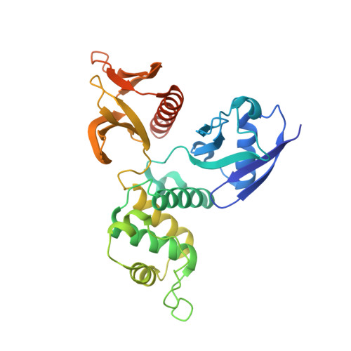
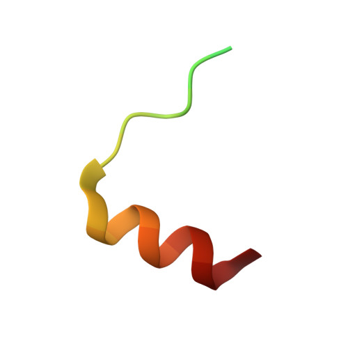
    The Na+/H+ exchanger regulatory factor (NHERF) is a key adaptor protein involved in the anchoring of ion channels and receptors to the actin cytoskeleton through binding to ERM (ezrin/radixin/moesin) proteins. NHERF binds the FERM domain of ERM proteins, although NHERF has no signature Motif-1 sequence for FERM binding found in adhesion molecules. The crystal structures of the radixin FERM domain complexed with the NHERF-1 and NHERF-2 C-terminal peptides revealed a peptide binding site of the FERM domain specific for the 13 residue motif MDWxxxxx(L/I)Fxx(L/F) (Motif-2), which is distinct from Motif-1. This Motif-2 forms an amphipathic alpha helix for hydrophobic docking to subdomain C of the FERM domain. This docking causes induced-fit conformational changes in subdomain C and affects binding to adhesion molecule peptides, while the two binding sites are not overlapped. Our studies provide structural paradigms for versatile ERM linkages between membrane proteins and the cytoskeleton.


  • Organizational Affiliation

    Structural Biology Laboratory, Nara Institute of Science and Technology, 8916-5 Takayama, Ikoma, Nara 630-0192, Japan.


Macromolecules
Find similar proteins by:  (by identity cutoff)  |  3D Structure
Entity ID: 1
MoleculeChains Sequence LengthOrganismDetailsImage
Radixin
A, B, C, D
312Mus musculusMutation(s): 0 
UniProt & NIH Common Fund Data Resources
Find proteins for P26043 (Mus musculus)
Explore P26043 
Go to UniProtKB:  P26043
IMPC:  MGI:97887
Entity Groups  
Sequence Clusters30% Identity50% Identity70% Identity90% Identity95% Identity100% Identity
UniProt GroupP26043
Sequence Annotations
Expand
  • Reference Sequence
Find similar proteins by:  (by identity cutoff)  |  3D Structure
Entity ID: 2
MoleculeChains Sequence LengthOrganismDetailsImage
Na(+)/H(+) exchange regulatory cofactor NHE-RF2
E, F, G, H
28N/AMutation(s): 0 
UniProt & NIH Common Fund Data Resources
Find proteins for Q15599 (Homo sapiens)
Explore Q15599 
Go to UniProtKB:  Q15599
PHAROS:  Q15599
GTEx:  ENSG00000065054 
Entity Groups  
Sequence Clusters30% Identity50% Identity70% Identity90% Identity95% Identity100% Identity
UniProt GroupQ15599
Sequence Annotations
Expand
  • Reference Sequence
Experimental Data & Validation

Experimental Data

  • Method: X-RAY DIFFRACTION
  • Resolution: 2.81 Å
  • R-Value Free: 0.278 
  • R-Value Work: 0.222 
  • R-Value Observed: 0.223 
  • Space Group: P 21 21 21
Unit Cell:
Length ( Å )Angle ( ˚ )
a = 68.626α = 90
b = 144.375β = 90
c = 177.944γ = 90
Software Package:
Software NamePurpose
REFMACrefinement
DENZOdata reduction
SCALEPACKdata scaling
AMoREphasing

Structure Validation

View Full Validation Report


Entry History 

Deposition Data

Revision History  (Full details and data files)

  • Version 1.0: 2006-07-18
    Type: Initial release
  • Version 1.1: 2008-04-30
    Changes: Version format compliance
  • Version 1.2: 2011-07-13
    Changes: Version format compliance
  • Version 1.3: 2024-03-13
    Changes: Data collection, Database references