2D03 | pdb_00002d03

Crystal structure of the G91S mutant of the NNA7 Fab


Experimental Data Snapshot

  • Method: X-RAY DIFFRACTION
  • Resolution: 1.97 Å
  • R-Value Free: 
    0.262 (Depositor), 0.263 (DCC) 
  • R-Value Work: 
    0.218 (Depositor), 0.219 (DCC) 
  • R-Value Observed: 
    0.218 (Depositor) 

Starting Model: experimental
View more details

wwPDB Validation 3D Report Full Report

Validation slider image for 2D03

This is version 1.4 of the entry. See complete history

Literature

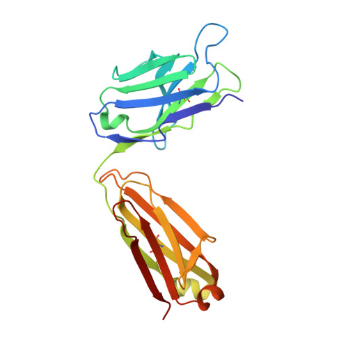
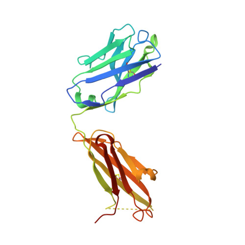
Crystallographic analysis of the NNA7 Fab and proposal for the mode of human blood-group recognition.

Xie, K.Song, S.C.Spitalnik, S.L.Wedekind, J.E.

(2005) Acta Crystallogr D Biol Crystallogr 61: 1386-1394

  • DOI: https://doi.org/10.1107/S0907444905023851
  • Primary Citation Related Structures: 
    2D03

  • PubMed Abstract: 

    The NNA7 Fab antibody fragment recognizes the human N-type blood-group antigen comprised of the N-terminal glycopeptide of glycophorin A (GPA). A mutant form of this Fab fragment, NNA7-G91S, exhibits markedly reduced antigen binding. To provide insight into how these Fab fragments recognize this glycopeptide antigen, the crystal structures of NNA7 and NNA7-G91S were solved and refined to 1.83 and 1.97 A resolution, respectively. Both molecules are composed of the same heavy (H) chain Fd fragment, but each contains a slightly different light (L) chain owing to the G91S substitution. Specifically, the G91S mutation pushes the backbone of the neighboring H chain away from complementarity-determining region 3 (CDR3) of the L-chain variable region, allowing an additional glycerol cryoprotectant molecule to enter the antigen-combining site near the putative location of O-linked glycosylation. Each Fab fragment also possesses a well defined 2-(N-morpholino)ethanesulfonic acid (MES) molecule trapped in its antigen-combining site, as well as a crystallographic symmetry-related molecule comprising an amino-acid sequence that is virtually identical to the N-terminus of GPA. The MES molecule interacts with the H-chain CDR in a manner reminiscent of antibody-carbohydrate complexes. These results suggest a model for recognition of the glycopeptide antigen that accounts for the deleterious effect of the G91S substitution.


  • Organizational Affiliation
    • Department of Biochemistry and Biophysics, University of Rochester School of Medicine and Dentistry, Rochester, NY 14642, USA.

Macromolecule Content 

  • Total Structure Weight: 48.61 kDa 
  • Atom Count: 3,639 
  • Modeled Residue Count: 429 
  • Deposited Residue Count: 440 
  • Unique protein chains: 2

Macromolecules

Find similar proteins by:|  3D Structure
Entity ID: 1
MoleculeChains  Sequence LengthOrganismDetailsImage
anti-glycophorin A type N immunoglobulin light chainA [auth L]217Mus musculusMutation(s): 0 
Find similar proteins by:|  3D Structure
Entity ID: 2
MoleculeChains  Sequence LengthOrganismDetailsImage
anti-glycophorin A type N immunoglobulin heavy chainB [auth H]223Mus musculusMutation(s): 0 

Small Molecules

Ligands 3 Unique
IDChains Name / Formula / InChI Key2D Diagram3D Interactions
MES

Query on MES


Download:Ideal Coordinates CCD File
H
2-(N-MORPHOLINO)-ETHANESULFONIC ACID
C6 H13 N O4 S
SXGZJKUKBWWHRA-UHFFFAOYSA-N
PEG

Query on PEG


Download:Ideal Coordinates CCD File
K [auth H]DI(HYDROXYETHYL)ETHER
C4 H10 O3
MTHSVFCYNBDYFN-UHFFFAOYSA-N
GOL

Query on GOL


Download:Ideal Coordinates CCD File
C [auth L]
D [auth L]
E [auth L]
F [auth L]
G [auth L]
C [auth L],
D [auth L],
E [auth L],
F [auth L],
G [auth L],
I [auth H],
J [auth H]
GLYCEROL
C3 H8 O3
PEDCQBHIVMGVHV-UHFFFAOYSA-N

Experimental Data & Validation

Experimental Data

  • Method: X-RAY DIFFRACTION
  • Resolution: 1.97 Å
  • R-Value Free:  0.262 (Depositor), 0.263 (DCC) 
  • R-Value Work:  0.218 (Depositor), 0.219 (DCC) 
  • R-Value Observed: 0.218 (Depositor) 
Space Group: P 21 21 21
Unit Cell:
Length ( Å )Angle ( ˚ )
a = 57.56α = 90
b = 70.65β = 90
c = 122.52γ = 90
Software Package:
Software NamePurpose
CNSrefinement
HKL-2000data reduction
CrystalCleardata scaling
AMoREphasing

Structure Validation

View Full Validation Report


Entry History 

Deposition Data

Revision History  (Full details and data files)

  • Version 1.0: 2006-01-24
    Type: Initial release
  • Version 1.1: 2008-04-30
    Changes: Version format compliance
  • Version 1.2: 2011-07-13
    Changes: Non-polymer description, Version format compliance
  • Version 1.3: 2023-10-25
    Changes: Data collection, Database references, Derived calculations, Refinement description
  • Version 1.4: 2024-11-13
    Changes: Structure summary