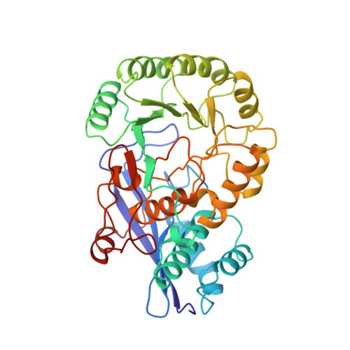
A re-evaluation of the crystal structure of chloromuconate cycloisomerase.
Kleywegt, G.J., Hoier, H., Jones, T.A.(1996) Acta Crystallogr D Biol Crystallogr 52: 858-863
- PubMed: 15299651 Search on PubMed
- DOI: https://doi.org/10.1107/S0907444995008936
- Primary Citation Related Structures:
2CHR - PubMed Abstract:
It is shown here that the reported 3 A crystal structure of chloromuconate cycloisomerase from Alcaligenes eutrophus [Hoier, Schlömann, Hammer, Glusker, Carrell, Goldman, Stezowski & Heinemann (1994). Acta Cryst. D50, 75-84] was refined in the incorrect space group I4. In addition, a stretch of about 25 residues near the N-terminus is out-of-register with the density in the original structure. From the coordinates and structure factors deposited in the Protein Data Bank (PDB), it was possible to determine the correct space group to be I422. The structure was then re-refined, using the original data reduced to I422, to a crystallographic free R factor of 0.264 at 3 A resolution (conventional R factor 0.189). With conservative refinement and rebuilding methods, the errors in the chain tracing could be identified and remedied. Since the two molecules per asymmetric unit in the original structure are actually related by crystallographic symmetry, the observed differences between them are artefacts. In particular, the differences between, and peculiarities of the metal-binding sites are unreal. This case shows the dangers of crystallographic refinement in cases with unfavourable data-to-parameter ratios, and the importance of reducing the number of parameters in such cases to prevent gross errors (for instance, by using NCS constraints). It also demonstrates how the evaluation and monitoring of model quality during the entire refinement and rebuilding process can be used to detect and remedy serious errors. Finally, it presents a strong case in favour of depositing not only model coordinates, but also experimental data (preferably, both merged and unmerged data).
- Department of Molecular Biology, Biomedical Centre, Uppsala University, Sweden.
Organizational Affiliation: