2CHG | pdb_00002chg

Replication Factor C domains 1 and 2


Experimental Data Snapshot

  • Method: X-RAY DIFFRACTION
  • Resolution: 2.10 Å
  • R-Value Free: 
    0.256 (Depositor), 0.253 (DCC) 
  • R-Value Work: 
    0.212 (Depositor) 
  • R-Value Observed: 
    0.214 (Depositor) 

wwPDB Validation 3D Report Full Report

Validation slider image for 2CHG

Ligand Structure Quality Assessment 


This is version 1.3 of the entry. See complete history

Literature

Communication between Subunits within an Archaeal Clamp-Loader Complex.

Seybert, A.Singleton, M.R.Cook, N.Hall, D.R.Wigley, D.B.

(2006) EMBO J 25: 2209

  • DOI: https://doi.org/10.1038/sj.emboj.7601093
  • Primary Citation Related Structures: 
    2CHG, 2CHQ, 2CHV

  • PubMed Abstract: 

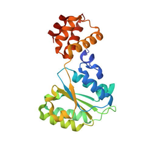
    We have investigated the communication between subunits in replication factor C (RFC) from Archaeoglobus fulgidus. Mutation of the proposed arginine finger in the small subunits results in a complex that can still bind ATP but has impaired clamp-loading activity, a process that normally only requires binding of nucleotide. The small subunit alone forms a hexameric ring that is six-fold symmetric in the absence of ATP. However, this symmetry is broken when the nucleotide is bound to the complex. A conformational change associated with nucleotide binding may relate to the opening of PCNA rings by RFC during the loading reaction. The structures also reveal the importance of the N-terminal helix of each subunit at the ATP-binding site. Analysis of mutant protein complexes containing subunits lacking this N-terminal helix reveals key distinct regulatory roles during clamp loading that are different for the large and small subunits in the RFC complex.


  • Organizational Affiliation
    • Clare Hall Laboratories, Cancer Research UK, London Research Institute, South Mimms Potters Bar, Herts, UK.

Macromolecule Content 

  • Total Structure Weight: 103.64 kDa 
  • Atom Count: 7,338 
  • Modeled Residue Count: 887 
  • Deposited Residue Count: 904 
  • Unique protein chains: 1

Macromolecules

Find similar proteins by:|  3D Structure
Entity ID: 1
MoleculeChains  Sequence LengthOrganismDetailsImage
REPLICATION FACTOR C SMALL SUBUNIT
A, B, C, D
226Archaeoglobus fulgidusMutation(s): 0 
UniProt
Find proteins for O28219 (Archaeoglobus fulgidus (strain ATCC 49558 / DSM 4304 / JCM 9628 / NBRC 100126 / VC-16))
Explore O28219 
Go to UniProtKB:  O28219
Entity Groups
Sequence Clusters30% Identity50% Identity70% Identity90% Identity95% Identity100% Identity
UniProt GroupO28219
Sequence Annotations
Expand
Reference Sequence

Small Molecules

Ligands 2 Unique
IDChains Name / Formula / InChI Key2D Diagram3D Interactions
ANP

Query on ANP


Download:Ideal Coordinates CCD File
E [auth A],
G [auth B],
I [auth C],
K [auth D]
PHOSPHOAMINOPHOSPHONIC ACID-ADENYLATE ESTER
C10 H17 N6 O12 P3
PVKSNHVPLWYQGJ-KQYNXXCUSA-N
MG

Query on MG


Download:Ideal Coordinates CCD File
F [auth A],
H [auth B],
J [auth C],
L [auth D]
MAGNESIUM ION
Mg
JLVVSXFLKOJNIY-UHFFFAOYSA-N

Experimental Data & Validation

Experimental Data

  • Method: X-RAY DIFFRACTION
  • Resolution: 2.10 Å
  • R-Value Free:  0.256 (Depositor), 0.253 (DCC) 
  • R-Value Work:  0.212 (Depositor) 
  • R-Value Observed: 0.214 (Depositor) 
Space Group: P 21 21 21
Unit Cell:
Length ( Å )Angle ( ˚ )
a = 77.504α = 90
b = 91.472β = 90
c = 140.644γ = 90
Software Package:
Software NamePurpose
REFMACrefinement

Structure Validation

View Full Validation Report


Ligand Structure Quality Assessment 


Entry History 

Deposition Data

Revision History  (Full details and data files)

  • Version 1.0: 2006-06-06
    Type: Initial release
  • Version 1.1: 2011-05-08
    Changes: Version format compliance
  • Version 1.2: 2011-07-13
    Changes: Version format compliance
  • Version 1.3: 2024-05-08
    Changes: Data collection, Database references, Derived calculations, Other