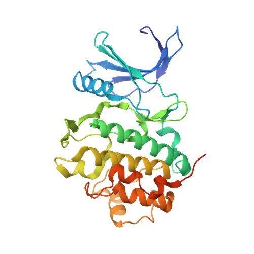
2CGU

Identification of chemically diverse Chk1 inhibitors by receptor- based virtual screening


Experimental Data Snapshot

  • Method: X-RAY DIFFRACTION
  • Resolution: 2.50 Å
  • R-Value Free: 0.271 
  • R-Value Work: 0.181 
  • R-Value Observed: 0.185 

Starting Model: experimental
View more details

wwPDB Validation   3D Report Full Report


This is version 1.3 of the entry. See complete history


Literature

Identification of Chemically Diverse Chk1 Inhibitors by Receptor-Based Virtual Screening.

Foloppe, N.Fisher, L.M.Howes, R.Potter, A.Robertson, A.G.S.Surgenor, A.E.

(2006) Bioorg Med Chem 14: 4792

  • DOI: https://doi.org/10.1016/j.bmc.2006.03.021
  • Primary Citation of Related Structures:  
    2CGU, 2CGV, 2CGW, 2CGX

  • PubMed Abstract: 

    Inhibition of the Chk1 kinase by small molecules is of great therapeutic interest for oncology and in understanding the cellular regulation of the G2/M checkpoint. We report how computational docking of a large electronic catalogue of compounds to an X-ray structure of the Chk1 ATP-binding site allowed prioritisation of a small subset of these compounds for assay. This led to the discovery of 10 novel Chk1 inhibitors, distributed among nine new and clearly different chemical scaffolds. Several of these scaffolds have promising lead-like properties. All these ligands act by competitive binding to the targeted ATP site. The crystal structures of four of these compounds bound to this site are presented, and reasonable modelled docking modes are suggested for the 5 other scaffolds. This structural context is used to assess the potential of these scaffolds for further medicinal chemistry efforts, suggesting that several of them could be elaborated to make additional interactions with the buried part of the ATP site. Some unusual interactions with the conserved kinase backbone motif are pointed out. The ligand-binding modes are also used to discuss their medicinal chemistry potential with respect to undesirable chemical functionalities, whether these functionalities bind directly to the protein or not. Overall, this work illustrates how virtual screening can identify a diverse set of ligands which bind to the targeted site. The structural models for these ligands in the Chk1 ATP-binding site will facilitate further medicinal chemistry efforts targeting this kinase.


  • Organizational Affiliation

    Vernalis (R&D) Ltd, Granta Park, Abington, Cambridge CB1 6GB, UK. n.foloppe@vernalis.com


Macromolecules
Find similar proteins by:  (by identity cutoff)  |  3D Structure
Entity ID: 1
MoleculeChains Sequence LengthOrganismDetailsImage
SERINE/THREONINE-PROTEIN KINASE CHK1297Homo sapiensMutation(s): 0 
EC: 2.7.1.37
UniProt & NIH Common Fund Data Resources
Find proteins for O14757 (Homo sapiens)
Explore O14757 
Go to UniProtKB:  O14757
PHAROS:  O14757
GTEx:  ENSG00000149554 
Entity Groups  
Sequence Clusters30% Identity50% Identity70% Identity90% Identity95% Identity100% Identity
UniProt GroupO14757
Sequence Annotations
Expand
  • Reference Sequence
Small Molecules
Ligands 1 Unique
IDChains Name / Formula / InChI Key2D Diagram3D Interactions
3A3
Query on 3A3

Download Ideal Coordinates CCD File 
B [auth A]2,2'-{[9-(HYDROXYIMINO)-9H-FLUORENE-2,7-DIYL]BIS(OXY)}DIACETIC ACID
C17 H13 N O7
VOKATEXROYSXDW-UHFFFAOYSA-N
Binding Affinity Annotations 
IDSourceBinding Affinity
3A3 PDBBind:  2CGU IC50: 1.34e+4 (nM) from 1 assay(s)
Experimental Data & Validation

Experimental Data

  • Method: X-RAY DIFFRACTION
  • Resolution: 2.50 Å
  • R-Value Free: 0.271 
  • R-Value Work: 0.181 
  • R-Value Observed: 0.185 
  • Space Group: P 1 21 1
Unit Cell:
Length ( Å )Angle ( ˚ )
a = 44.993α = 90
b = 65.787β = 102.28
c = 54.625γ = 90
Software Package:
Software NamePurpose
REFMACrefinement
DENZOdata reduction
SCALEPACKdata scaling
AMoREphasing

Structure Validation

View Full Validation Report


Entry History 

Deposition Data

Revision History  (Full details and data files)

  • Version 1.0: 2006-04-05
    Type: Initial release
  • Version 1.1: 2011-05-08
    Changes: Version format compliance
  • Version 1.2: 2011-07-13
    Changes: Version format compliance
  • Version 1.3: 2023-12-13
    Changes: Data collection, Database references, Other, Refinement description