


Crystal Structure of Human Pyridoxal 5'-Phosphate Phosphatase
Kang, B.S., Cho, H.J., Kim, K.J., Kwon, O.S.To be published.
Experimental Data Snapshot
wwPDB Validation 3D Report Full Report

Entity ID: 1 | |||||
|---|---|---|---|---|---|
| Molecule | Chains | Sequence Length | Organism | Details | Image |
| PYRIDOXAL PHOSPHATE PHOSPHATASE | 298 | Homo sapiens | Mutation(s): 0 EC: 3.1.3.74 (PDB Primary Data), 3.1.3.16 (UniProt) | 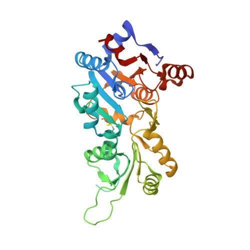 | |
UniProt & NIH Common Fund Data Resources | |||||
PHAROS: Q96GD0 GTEx: ENSG00000241360 | |||||
Entity Groups | |||||
| Sequence Clusters | 30% Identity50% Identity70% Identity90% Identity95% Identity100% Identity | ||||
| UniProt Group | Q96GD0 | ||||
Sequence AnnotationsExpand | |||||
Reference Sequence | |||||
| Ligands 1 Unique | |||||
|---|---|---|---|---|---|
| ID | Chains | Name / Formula / InChI Key | 2D Diagram | 3D Interactions | |
| MG Download:Ideal Coordinates CCD File | B [auth A], C [auth A] | MAGNESIUM ION Mg JLVVSXFLKOJNIY-UHFFFAOYSA-N |  | ||
| Length ( Å ) | Angle ( ˚ ) |
|---|---|
| a = 54.631 | α = 90 |
| b = 54.631 | β = 90 |
| c = 213.985 | γ = 90 |
| Software Name | Purpose |
|---|---|
| REFMAC | refinement |


