2CEU | pdb_00002ceu

Despentapeptide insulin in acetic acid (pH 2)


Experimental Data Snapshot

  • Method: X-RAY DIFFRACTION
  • Resolution: 1.80 Å
  • R-Value Free: 
    0.211 (Depositor), 0.204 (DCC) 
  • R-Value Work: 
    0.172 (Depositor), 0.180 (DCC) 
  • R-Value Observed: 
    0.174 (Depositor) 

Starting Model: other
View more details

wwPDB Validation 3D Report Full Report

Validation slider image for 2CEU

This is version 1.4 of the entry. See complete history

Literature

I222 Crystal Form of Despentapeptide (B26-B30) Insulin Provides New Insights Into the Properties of Monomeric Insulin.

Whittingham, J.L.Youshang, Z.Zakova, L.Dodson, E.J.Turkenburg, J.P.Brange, J.Dodson, G.G.

(2006) Acta Crystallogr D Biol Crystallogr 62: 505

  • DOI: https://doi.org/10.1107/S0907444906006871
  • Primary Citation Related Structures: 
    2CEU

  • PubMed Abstract: 

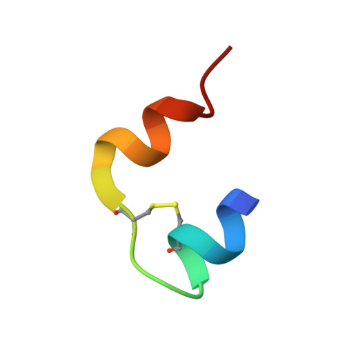
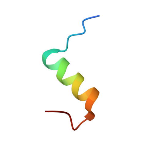
    Despentapeptide (des-B26-B30) insulin (DPI), an active modified insulin, has been crystallized in the presence of 20% acetic acid pH 2. A crystal structure analysis to 1.8 A spacing (space group I222) revealed that the DPI molecule, which is unable to make beta-strand interactions for physiological dimer formation and is apparently monomeric in solution, formed an alternative lattice-generated dimer. The formation of this dimer involved interactions between surfaces which included the B9-B19 alpha-helices (usually buried by the dimer-dimer contacts within the native hexamer). The two crystallographically independent molecules within the dimer were essentially identical and were similar in conformation to T-state insulin as seen in the T(6) insulin hexamer. An unusual feature of each molecule in the dimer was the presence of two independent conformations at the B-chain C-terminus (residues B20-B25). Both conformations were different from that of native insulin, involving a 3.5 A displacement of the B20-B23 beta-turn and a repositioning of residue PheB25 such that it made close van der Waals contact with the main body of the molecule, appearing to stabilize the B-chain C-terminus.


  • Organizational Affiliation
    • York Structural Biology Laboratory, Department of Chemistry, University of York, Heslington, York YO10 5YW, England.

Macromolecule Content 

  • Total Structure Weight: 10.74 kDa 
  • Atom Count: 854 
  • Modeled Residue Count: 90 
  • Deposited Residue Count: 92 
  • Unique protein chains: 2

Macromolecules

Find similar proteins by:  Sequence   |   3D Structure  
Entity ID: 1
MoleculeChains  Sequence LengthOrganismDetailsImage
INSULIN
A, C
21Homo sapiensMutation(s): 0 
UniProt & NIH Common Fund Data Resources
Find proteins for P01308 (Homo sapiens)
Explore P01308 
Go to UniProtKB:  P01308
PHAROS:  P01308
GTEx:  ENSG00000254647 
Entity Groups
Sequence Clusters30% Identity50% Identity70% Identity90% Identity95% Identity100% Identity
UniProt GroupP01308
Sequence Annotations
Expand
Reference Sequence
Find similar proteins by:  Sequence   |   3D Structure  
Entity ID: 2
MoleculeChains  Sequence LengthOrganismDetailsImage
INSULIN
B, D
25Homo sapiensMutation(s): 0 
UniProt & NIH Common Fund Data Resources
Find proteins for P01308 (Homo sapiens)
Explore P01308 
Go to UniProtKB:  P01308
PHAROS:  P01308
GTEx:  ENSG00000254647 
Entity Groups
Sequence Clusters30% Identity50% Identity70% Identity90% Identity95% Identity100% Identity
UniProt GroupP01308
Sequence Annotations
Expand
Reference Sequence

Experimental Data & Validation

Experimental Data

  • Method: X-RAY DIFFRACTION
  • Resolution: 1.80 Å
  • R-Value Free:  0.211 (Depositor), 0.204 (DCC) 
  • R-Value Work:  0.172 (Depositor), 0.180 (DCC) 
  • R-Value Observed: 0.174 (Depositor) 
Space Group: I 2 2 2
Unit Cell:
Length ( Å )Angle ( ˚ )
a = 58.152α = 90
b = 58.174β = 90
c = 54.797γ = 90
Software Package:
Software NamePurpose
REFMACrefinement
DENZOdata reduction
SCALEPACKdata scaling
AMoREphasing

Structure Validation

View Full Validation Report


Entry History 

Deposition Data

Revision History  (Full details and data files)

  • Version 1.0: 2006-03-03
    Type: Initial release
  • Version 1.1: 2011-05-08
    Changes: Version format compliance
  • Version 1.2: 2011-07-13
    Changes: Version format compliance
  • Version 1.3: 2024-05-01
    Changes: Data collection, Database references, Derived calculations, Other, Refinement description
  • Version 1.4: 2024-11-20
    Changes: Structure summary