2C9S | pdb_00002c9s

1.24 Angstroms resolution structure of Zn-Zn Human Superoxide dismutase


Experimental Data Snapshot

  • Method: X-RAY DIFFRACTION
  • Resolution: 1.24 Å
  • R-Value Free: 
    0.194 (Depositor), 0.204 (DCC) 
  • R-Value Work: 
    0.152 (Depositor), 0.162 (DCC) 
  • R-Value Observed: 
    0.154 (Depositor) 

Starting Model: experimental
View more details

wwPDB Validation 3D Report Full Report

Validation slider image for 2C9S

This is version 1.4 of the entry. See complete history

Literature

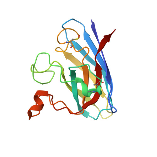
Variable Metallation of Human Superoxide Dismutase: Atomic Resolution Crystal Structures of Cu-Zn, Zn-Zn and as-Isolated Wild-Type Enzymes.

Strange, R.W.Antonyuk, S.V.Hough, M.A.Doucette, P.A.Valentine, J.S.Hasnain, S.S.

(2006) J Mol Biology 356: 1152

  • DOI: https://doi.org/10.1016/j.jmb.2005.11.081
  • Primary Citation Related Structures: 
    2C9S, 2C9U, 2C9V

  • PubMed Abstract: 

    Human Cu-Zn superoxide dismutase (SOD1) protects cells from the effects of oxidative stress. Mutations in SOD1 are linked to the familial form of amyotrophic lateral sclerosis. Several hypotheses for their toxicity involve the mis-metallation of the enzyme. We present atomic-resolution crystal structures and biophysical data for human SOD1 in three metallation states: Zn-Zn, Cu-Zn and as-isolated. These data represent the first atomic-resolution structures for human SOD1, the first structure of a reduced SOD1, and the first structure of a fully Zn-substituted SOD1 enzyme. Recombinantly expressed as-isolated SOD1 contains a mixture of Zn and Cu at the Cu-binding site. The Zn-Zn structure appears to be at least as stable as the correctly (Cu-Zn) metallated enzyme. These data raise the possibility that in a cellular environment with low availability of free copper, Zn-Zn may be the preferred metallation state of SOD1 prior to its interaction with the copper chaperone.


  • Organizational Affiliation
    • Molecular Biophysics Group, CCLRC Daresbury Laboratory, Warrington, Cheshire WA4 4AD, UK.

Macromolecule Content 

  • Total Structure Weight: 32.83 kDa 
  • Atom Count: 2,966 
  • Modeled Residue Count: 306 
  • Deposited Residue Count: 306 
  • Unique protein chains: 1

Macromolecules

Find similar proteins by:|  3D Structure
Entity ID: 1
MoleculeChains  Sequence LengthOrganismDetailsImage
SUPEROXIDE DISMUTASE [CU-ZN]A,
B [auth F]
153Homo sapiensMutation(s): 0 
EC: 1.15.1.1 (PDB Primary Data), 1.8 (UniProt)
UniProt & NIH Common Fund Data Resources
Find proteins for P00441 (Homo sapiens)
Explore P00441 
Go to UniProtKB:  P00441
PHAROS:  P00441
GTEx:  ENSG00000142168 
Entity Groups
Sequence Clusters30% Identity50% Identity70% Identity90% Identity95% Identity100% Identity
UniProt GroupP00441
Sequence Annotations
Expand
Reference Sequence

Small Molecules

Ligands 3 Unique
IDChains Name / Formula / InChI Key2D Diagram3D Interactions
SO4

Query on SO4


Download:Ideal Coordinates CCD File
H [auth A]
I [auth A]
J [auth A]
K [auth A]
L [auth F]
H [auth A],
I [auth A],
J [auth A],
K [auth A],
L [auth F],
M [auth F]
SULFATE ION
O4 S
QAOWNCQODCNURD-UHFFFAOYSA-L
ZN

Query on ZN


Download:Ideal Coordinates CCD File
C [auth A]
D [auth A]
E [auth A]
N [auth F]
O [auth F]
C [auth A],
D [auth A],
E [auth A],
N [auth F],
O [auth F],
Q [auth F]
ZINC ION
Zn
PTFCDOFLOPIGGS-UHFFFAOYSA-N
ACT

Query on ACT


Download:Ideal Coordinates CCD File
F [auth A],
G [auth A],
P [auth F]
ACETATE ION
C2 H3 O2
QTBSBXVTEAMEQO-UHFFFAOYSA-M
Modified Residues  1 Unique
IDChains TypeFormula2D DiagramParent
CSO
Query on CSO
A,
B [auth F]
L-PEPTIDE LINKINGC3 H7 N O3 SCYS

Experimental Data & Validation

Experimental Data

  • Method: X-RAY DIFFRACTION
  • Resolution: 1.24 Å
  • R-Value Free:  0.194 (Depositor), 0.204 (DCC) 
  • R-Value Work:  0.152 (Depositor), 0.162 (DCC) 
  • R-Value Observed: 0.154 (Depositor) 
Space Group: P 1 21 1
Unit Cell:
Length ( Å )Angle ( ˚ )
a = 38.243α = 90
b = 67.742β = 106.44
c = 51.26γ = 90
Software Package:
Software NamePurpose
REFMACrefinement
HKL-2000data reduction
HKL-2000data scaling
MOLREPphasing

Structure Validation

View Full Validation Report


Entry History 

Deposition Data

Revision History  (Full details and data files)

  • Version 1.0: 2005-12-15
    Type: Initial release
  • Version 1.1: 2011-05-08
    Changes: Version format compliance
  • Version 1.2: 2011-07-13
    Changes: Version format compliance
  • Version 1.3: 2023-12-13
    Changes: Data collection, Database references, Derived calculations, Other, Refinement description
  • Version 1.4: 2024-11-20
    Changes: Structure summary