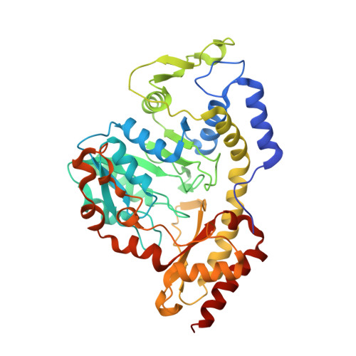
Crystal structures of the PLP- and PMP-bound forms of BtrR, a dual functional aminotransferase involved in butirosin biosynthesis.
Popovic, B., Tang, X., Chirgadze, D.Y., Huang, F., Blundell, T.L., Spencer, J.B.(2006) Proteins 65: 220-230
- PubMed: 16894611 Search on PubMed
- DOI: https://doi.org/10.1002/prot.21076
- Primary Citation Related Structures:
2C7T, 2C81 - PubMed Abstract:
The aminotransferase (BtrR), which is involved in the biosynthesis of butirosin, a 2-deoxystreptamine (2-DOS)-containing aminoglycoside antibiotic produced by Bacillus circulans, catalyses the pyridoxal phosphate (PLP)-dependent transamination reaction both of 2-deoxy-scyllo-inosose to 2-deoxy-scyllo-inosamine and of amino-dideoxy-scyllo-inosose to 2-DOS. The high-resolution crystal structures of the PLP- and PMP-bound forms of BtrR aminotransferase from B. circulans were solved at resolutions of 2.1 A and 1.7 A with R(factor)/R(free) values of 17.4/20.6 and 19.9/21.9, respectively. BtrR has a fold characteristic of the aspartate aminotransferase family, and sequence and structure analysis categorises it as a member of SMAT (secondary metabolite aminotransferases) subfamily. It exists as a homodimer with two active sites per dimer. The active site of the BtrR protomer is located in a cleft between an alpha helical N-terminus, a central alphabetaalpha sandwich domain and an alphabeta C-terminal domain. The structures of the PLP- and PMP-bound enzymes are very similar; however BtrR-PMP lacks the covalent bond to Lys192. Furthermore, the two forms differ in the side-chain conformations of Trp92, Asp163, and Tyr342 that are likely to be important in substrate selectivity and substrate binding. This is the first three-dimensional structure of an enzyme from the butirosin biosynthesis gene cluster.
- Department of Biochemistry, University of Cambridge, Cambridge, United Kingdom.
Organizational Affiliation: