2C6R | pdb_00002c6r

FE-SOAKED CRYSTAL STRUCTURE OF THE DPS92 FROM DEINOCOCCUS RADIODURANS


Experimental Data Snapshot

  • Method: X-RAY DIFFRACTION
  • Resolution: 2.10 Å
  • R-Value Work: 
    0.188 (Depositor), 0.185 (DCC) 
  • R-Value Observed: 
    0.188 (Depositor) 

wwPDB Validation 3D Report Full Report

Validation slider image for 2C6R

This is version 1.3 of the entry. See complete history

Literature

The Crystal Structure of the Dps2 from Deinococcus Radiodurans Reveals an Unusual Pore Profile with a Non-Specific Metal Binding Site.

Cuypers, M.G.Mitchell, E.P.Romao, C.V.Mcsweeney, S.M.

(2007) J Mol Biology 371: 787

  • DOI: https://doi.org/10.1016/j.jmb.2006.11.032
  • Primary Citation Related Structures: 
    2C2J, 2C6R

  • PubMed Abstract: 

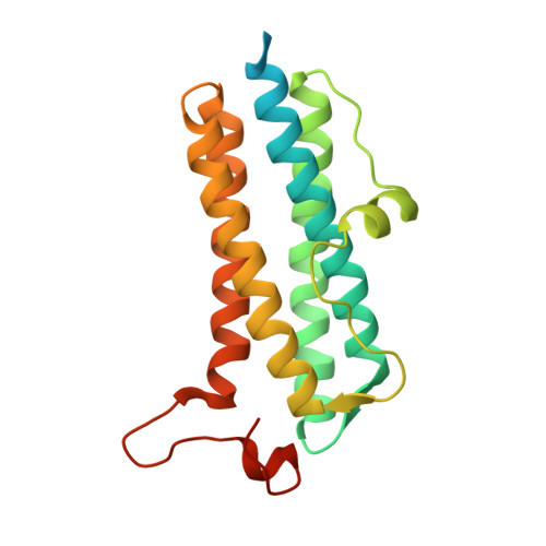
    The crystal structure of recombinant Dps2 (DRB0092, DNA protecting protein under starved conditions) from the Gram-positive, radiation-resistant bacterium Deinococcus radiodurans has been determined in its apo and iron loaded states. Like other members of the Dps family, the bacterial DrDps2 assembles as a spherical dodecamer with an outer shell diameter of 90 A and an interior diameter of 40 A. A total of five iron sites were located in the iron loaded structure, representing the first stages of iron biomineralisation. Each subunit contains a mononuclear iron ferroxidase centre coordinated by residues highly conserved amongst the Dps family of proteins. In the structures presented, a distinct iron site is observed 6.1 A from the ferroxidase centre with a unique ligand configuration of mono coordination by the protein and no bridging ligand to the ferroxidase centre. A non-specific metallic binding site, suspected to play a regulative role in iron uptake/release from the cage, was found in a pocket located near to the external edge of the C-terminal 3-fold channel.


  • Organizational Affiliation
    • ESRF (European Synchrotron Radiation Facility), 6 rue Jules Horowitz, BP 220, 38043 Grenoble, France.

Macromolecule Content 

  • Total Structure Weight: 23.6 kDa 
  • Atom Count: 1,474 
  • Modeled Residue Count: 166 
  • Deposited Residue Count: 211 
  • Unique protein chains: 1

Macromolecules

Find similar proteins by:|  3D Structure
Entity ID: 1
MoleculeChains  Sequence LengthOrganismDetailsImage
DNA-BINDING STRESS RESPONSE PROTEIN, DPS FAMILY211Deinococcus radiodurans R1 = ATCC 13939 = DSM 20539Mutation(s): 0 
EC: 1.16
UniProt
Find proteins for Q9RZN1 (Deinococcus radiodurans (strain ATCC 13939 / DSM 20539 / JCM 16871 / CCUG 27074 / LMG 4051 / NBRC 15346 / NCIMB 9279 / VKM B-1422 / R1))
Explore Q9RZN1 
Go to UniProtKB:  Q9RZN1
Entity Groups
Sequence Clusters30% Identity50% Identity70% Identity90% Identity95% Identity100% Identity
UniProt GroupQ9RZN1
Sequence Annotations
Expand
Reference Sequence

Experimental Data & Validation

Experimental Data

  • Method: X-RAY DIFFRACTION
  • Resolution: 2.10 Å
  • R-Value Work:  0.188 (Depositor), 0.185 (DCC) 
  • R-Value Observed: 0.188 (Depositor) 
Space Group: P 2 3
Unit Cell:
Length ( Å )Angle ( ˚ )
a = 88.965α = 90
b = 88.965β = 90
c = 88.965γ = 90
Software Package:
Software NamePurpose
REFMACrefinement
MOSFLMdata reduction
SCALAdata scaling

Structure Validation

View Full Validation Report


Entry History 

Deposition Data

Revision History  (Full details and data files)

  • Version 1.0: 2007-02-20
    Type: Initial release
  • Version 1.1: 2011-05-08
    Changes: Version format compliance
  • Version 1.2: 2011-07-13
    Changes: Version format compliance
  • Version 1.3: 2024-05-08
    Changes: Data collection, Database references, Derived calculations, Other