2C3K | pdb_00002c3k

Identification of a buried pocket for potent and selective inhibition of Chk1: prediction and verification


Experimental Data Snapshot

  • Method: X-RAY DIFFRACTION
  • Resolution: 2.60 Å
  • R-Value Free: 
    0.280 (Depositor), 0.268 (DCC) 
  • R-Value Work: 
    0.198 (Depositor), 0.192 (DCC) 
  • R-Value Observed: 
    0.202 (Depositor) 

Starting Model: experimental
View more details

wwPDB Validation 3D Report Full Report

Validation slider image for 2C3K

Ligand Structure Quality Assessment 


This is version 1.3 of the entry. See complete history

Literature

Identification of a Buried Pocket for Potent and Selective Inhibition of Chk1: Prediction and Verification.

Foloppe, N.Fisher, L.M.Francis, G.Howes, R.Kierstan, P.Potter, A.

(2006) Bioorg Med Chem 14: 1792

  • DOI: https://doi.org/10.1016/j.bmc.2005.10.022
  • Primary Citation Related Structures: 
    2C3J, 2C3K, 2C3L

  • PubMed Abstract: 

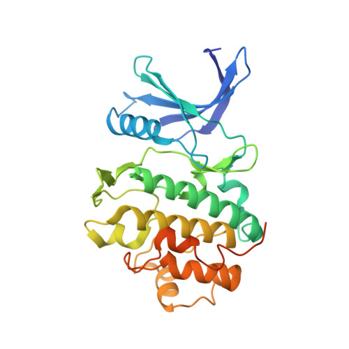
    Inhibition of the Chk1 kinase by small molecules binding to its active site is a strategy of great therapeutic interest for oncology. We report how computational modelling predicted the binding mode of ligands of special interest to the Chk1 ATP site, for representatives of an indazole series and debromohymenialdisine. These binding modes were subsequently confirmed by X-ray crystallography. The binding mode of a potent indazole derivative involves non-conventional C-H...O and N-H...pi-aromatic interactions with the protein. These interactions are formed in a buried pocket at the periphery of the ATP-binding site, the importance of which has previously been overlooked for ligand design against Chk1. It is demonstrated that filling this pocket can confer ligands with dramatically enhanced affinity for Chk1. Structural arguments in conjunction with assay data explain why targeting this pocket is also advantageous for selective binding to Chk1. Structural overlays of known inhibitors complexed with Chk1 show that only the indazole series utilizes the pocket of interest. Therefore, the analysis presented here should prove helpful in guiding future structure-based ligand design efforts against Chk1.


  • Organizational Affiliation
    • Vernalis (R&D) Ltd, Granta Park, Abington, Cambridge CB1 6GB, UK. nfoloppe@vernalis.com

Macromolecule Content 

  • Total Structure Weight: 34.5 kDa 
  • Atom Count: 2,272 
  • Modeled Residue Count: 262 
  • Deposited Residue Count: 297 
  • Unique protein chains: 1

Macromolecules

Find similar proteins by:|  3D Structure
Entity ID: 1
MoleculeChains  Sequence LengthOrganismDetailsImage
SERINE/THREONINE-PROTEIN KINASE CHK1297Homo sapiensMutation(s): 0 
EC: 2.7.1.37 (PDB Primary Data), 2.7.11.1 (UniProt)
UniProt & NIH Common Fund Data Resources
Find proteins for O14757 (Homo sapiens)
Explore O14757 
Go to UniProtKB:  O14757
PHAROS:  O14757
GTEx:  ENSG00000149554 
Entity Groups
Sequence Clusters30% Identity50% Identity70% Identity90% Identity95% Identity100% Identity
UniProt GroupO14757
Sequence Annotations
Expand
Reference Sequence

Small Molecules

Ligands 1 Unique
IDChains Name / Formula / InChI Key2D Diagram3D Interactions
ABO

Query on ABO


Download:Ideal Coordinates CCD File
B [auth A]4-[3-(1H-BENZIMIDAZOL-2-YL)-1H-INDAZOL-6-YL]-2-METHOXYPHENOL
C21 H16 N4 O2
GYPHGCGOSZJXOT-UHFFFAOYSA-N

Experimental Data & Validation

Experimental Data

  • Method: X-RAY DIFFRACTION
  • Resolution: 2.60 Å
  • R-Value Free:  0.280 (Depositor), 0.268 (DCC) 
  • R-Value Work:  0.198 (Depositor), 0.192 (DCC) 
  • R-Value Observed: 0.202 (Depositor) 
Space Group: P 1 21 1
Unit Cell:
Length ( Å )Angle ( ˚ )
a = 45.057α = 90
b = 65.891β = 102.01
c = 54.584γ = 90
Software Package:
Software NamePurpose
REFMACrefinement
HKL-2000data reduction
SCALEPACKdata scaling
AMoREphasing

Structure Validation

View Full Validation Report


Ligand Structure Quality Assessment 


Entry History 

Deposition Data

Revision History  (Full details and data files)

  • Version 1.0: 2005-11-23
    Type: Initial release
  • Version 1.1: 2011-05-07
    Changes: Version format compliance
  • Version 1.2: 2011-07-13
    Changes: Version format compliance
  • Version 1.3: 2023-12-13
    Changes: Data collection, Database references, Other, Refinement description