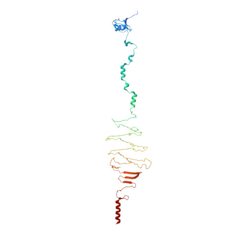
Structure of a Group a Streptococcal Phage-Encoded Virulence Factor Reveals Catalytically Active Triple-Stranded Beta-Helix
Smith, N.L., Taylor, E.J., Linsay, A.-M., Charnock, S.J., Turkenburg, J.P., Dodson, E.J., Davies, G.J., Black, G.W.(2005) Proc Natl Acad Sci U S A 102: 17652
- PubMed: 16314578
- DOI: https://doi.org/10.1073/pnas.0504782102
- Primary Citation of Related Structures:
2C3F - PubMed Abstract:
Streptococcus pyogenes (group A Streptococcus) causes severe invasive infections including scarlet fever, pharyngitis (streptococcal sore throat), skin infections, necrotizing fasciitis (flesh-eating disease), septicemia, erysipelas, cellulitis, acute rheumatic fever, and toxic shock. The conversion from nonpathogenic to toxigenic strains of S. pyogenes is frequently mediated by bacteriophage infection. One of the key bacteriophage-encoded virulence factors is a putative "hyaluronidase," HylP1, a phage tail-fiber protein responsible for the digestion of the S. pyogenes hyaluronan capsule during phage infection. Here we demonstrate that HylP1 is a hyaluronate lyase. The 3D structure, at 1.8-angstroms resolution, reveals an unusual triple-stranded beta-helical structure and provides insight into the structural basis for phage tail assembly and the role of phage tail proteins in virulence. Unlike the triple-stranded beta-helix assemblies of the bacteriophage T4 injection machinery and the tailspike endosialidase of the Escherichia coli K1 bacteriophage K1F, HylP1 possesses three copies of the active center on the triple-helical fiber itself without the need for an accessory catalytic domain. The triple-stranded beta-helix is not simply a structural scaffold, as previously envisaged; it is harnessed to provide a 200-angstroms-long substrate-binding groove for the optimal reduction in hyaluronan viscosity to aid phage penetration of the capsule.
- Chemical Biology Research Group, School of Applied Sciences, Northumbria University, Newcastle upon Tyne NE1 8ST, United Kingdom.
Organizational Affiliation:


