2C35 | pdb_00002c35

Subunits Rpb4 and Rpb7 of human RNA polymerase II


Experimental Data Snapshot

  • Method: X-RAY DIFFRACTION
  • Resolution: 2.70 Å
  • R-Value Free: 
    0.266 (Depositor), 0.251 (DCC) 
  • R-Value Work: 
    0.230 (Depositor), 0.221 (DCC) 
  • R-Value Observed: 
    0.230 (Depositor) 

Starting Model: experimental
View more details

wwPDB Validation 3D Report Full Report

Validation slider image for 2C35

This is version 1.4 of the entry. See complete history

Literature

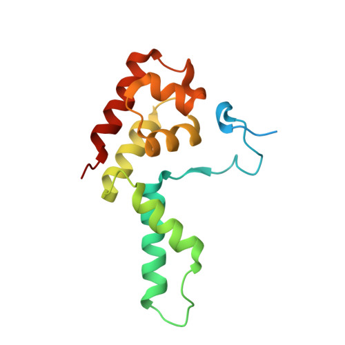
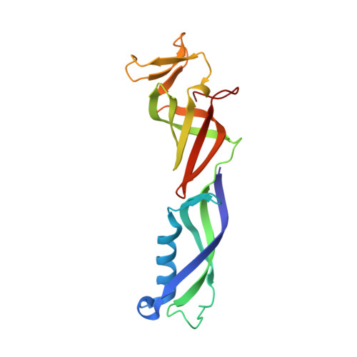
Crystal Structure and RNA Binding of the Rpb4/Rpb7 Subunits of Human RNA Polymerase II.

Meka, H.Werner, F.Cordell, S.C.Onesti, S.Brick, P.

(2005) Nucleic Acids Res 33: 6435

  • DOI: https://doi.org/10.1093/nar/gki945
  • Primary Citation Related Structures: 
    2C35

  • PubMed Abstract: 

    The Rpb4 and Rpb7 subunits of eukaryotic RNA polymerase II (RNAP(II)) form a heterodimer that protrudes from the 10-subunit core of the enzyme. We have obtained crystals of the human Rpb4/Rpb7 heterodimer and determined the structure to 2.7 A resolution. The presence of putative RNA-binding domains on the Rpb7 subunit and the position of the heterodimer close to the RNA exit groove in the 12 subunit yeast polymerase complex strongly suggests a role for the heterodimer in binding and stabilizing the nascent RNA transcript. We have complemented the structural analysis with biochemical studies directed at dissecting the RNA-binding properties of the human Rpb4/Rpb7 complex and that of the homologous E/F complex from Methanocaldococcus jannaschii. A number of conserved, solvent-exposed residues in both the human Rpb7 subunit and the archaeal E subunit have been modified by site-directed mutagenesis and the mutants tested for RNA binding by performing electrophoretic mobility shift assays. These studies have identified an elongated surface region on the corresponding face of both subunit E and Rpb7 that is involved in RNA binding. The area spans the nucleic acid binding face of the OB fold, including the B4-B5 loop, but also extends towards the N-terminal domain.


  • Organizational Affiliation
    • Department of Biological Sciences, Imperial College London, SW7 2BW, UK.

Macromolecule Content 

  • Total Structure Weight: 146.53 kDa 
  • Atom Count: 9,290 
  • Modeled Residue Count: 1,194 
  • Deposited Residue Count: 1,296 
  • Unique protein chains: 2

Macromolecules

Find similar proteins by:|  3D Structure
Entity ID: 1
MoleculeChains  Sequence LengthOrganismDetailsImage
DNA-DIRECTED RNA POLYMERASE II 16 KDA POLYPEPTIDE
A, C, E, G
152Homo sapiensMutation(s): 0 
EC: 2.7.7.6
UniProt & NIH Common Fund Data Resources
Find proteins for O15514 (Homo sapiens)
Explore O15514 
Go to UniProtKB:  O15514
PHAROS:  O15514
GTEx:  ENSG00000144231 
Entity Groups
Sequence Clusters30% Identity50% Identity70% Identity90% Identity95% Identity100% Identity
UniProt GroupO15514
Sequence Annotations
Expand
Reference Sequence
Find similar proteins by:|  3D Structure
Entity ID: 2
MoleculeChains  Sequence LengthOrganismDetailsImage
DNA-DIRECTED RNA POLYMERASE II 19 KDA POLYPEPTIDE
B, D, F, H
172Homo sapiensMutation(s): 0 
UniProt & NIH Common Fund Data Resources
Find proteins for P62487 (Homo sapiens)
Explore P62487 
Go to UniProtKB:  P62487
PHAROS:  P62487
GTEx:  ENSG00000168002 
Entity Groups
Sequence Clusters30% Identity50% Identity70% Identity90% Identity95% Identity100% Identity
UniProt GroupP62487
Sequence Annotations
Expand
Reference Sequence

Experimental Data & Validation

Experimental Data

  • Method: X-RAY DIFFRACTION
  • Resolution: 2.70 Å
  • R-Value Free:  0.266 (Depositor), 0.251 (DCC) 
  • R-Value Work:  0.230 (Depositor), 0.221 (DCC) 
  • R-Value Observed: 0.230 (Depositor) 
Space Group: P 1
Unit Cell:
Length ( Å )Angle ( ˚ )
a = 45.69α = 85.53
b = 83.19β = 81.8
c = 99.31γ = 81.77
Software Package:
Software NamePurpose
CNSrefinement
MOSFLMdata reduction
SCALAdata scaling
PHASERphasing

Structure Validation

View Full Validation Report


Entry History 

Deposition Data

Revision History  (Full details and data files)

  • Version 1.0: 2005-11-18
    Type: Initial release
  • Version 1.1: 2013-03-27
    Changes: Derived calculations, Non-polymer description, Other, Refinement description, Version format compliance
  • Version 1.2: 2019-03-06
    Changes: Data collection, Experimental preparation, Other
  • Version 1.3: 2019-05-08
    Changes: Data collection, Experimental preparation
  • Version 1.4: 2023-12-13
    Changes: Data collection, Database references, Other, Refinement description