Co-Axial Association of Recombinant Eye Lens Aquaporin-0 Observed in Loosely Packed 3D-Crystals
Palanivelu, D.V., Kozono, D.E., Engel, A., Suda, K., Lustig, A., Agre, P., Schirmer, T.(2006) J Mol Biology 355: 605
- PubMed: 16309700 Search on PubMed
- DOI: https://doi.org/10.1016/j.jmb.2005.10.032
- Primary Citation Related Structures:
2C32 - PubMed Abstract:
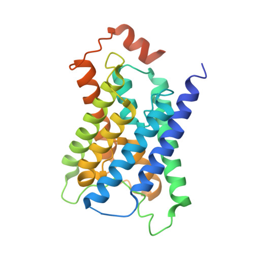
Aquaporin-0 (AQP0) is the major membrane protein in vertebrate eye lenses. It has been proposed that AQP0 tetramers mediate contact between membranes of adjacent lens fiber cells, which would be consistent with the extraordinarily narrow inter-cellular spacing. We have obtained 3D crystals of recombinant bovine AQP0 that diffract to 7.0 A resolution. The crystal packing was determined by molecular replacement and shows that, within the cubic lattice, AQP0 tetramers are associated head-to-head along their 4-fold axes. Oligomeric states larger than the tetramer were also observed in solution by native gel electrophoresis and analytical ultracentrifugation methods. In the crystals, there are no direct contacts between octamers, and it can thus be inferred that crystalline order is mediated solely by the detergent belts surrounding the membrane protein. Across the tetramer-tetramer interface, extracellular loops A and C interdigitate at the center and the perimeter of the octamer, respectively. The octamer structure is compared with that of the recently determined structure of truncated ovine AQP0 derived from electron diffraction of 2D crystals. Intriguingly, also in these crystals, octamers are observed, but with significantly different relative tetramer-tetramer orientations. The interactions observed in the loosely packed 3D crystals reported here may in fact represent an in vivo association mode between AQP0 tetramers from juxtaposed membranes in the eye lens.
- Division of Structural Biology, Biozentrum, University of Basel, Klingelbergstr. 70, CH-4056 Basel, Switzerland.
Organizational Affiliation: