2C2Z | pdb_00002c2z

Crystal structure of caspase-8 in complex with aza-peptide Michael acceptor inhibitor


Experimental Data Snapshot

  • Method: X-RAY DIFFRACTION
  • Resolution: 1.95 Å
  • R-Value Free: 
    0.187 (Depositor), 0.189 (DCC) 
  • R-Value Work: 
    0.171 (Depositor), 0.167 (DCC) 
  • R-Value Observed: 
    0.171 (Depositor) 

wwPDB Validation 3D Report Full Report

Validation slider image for 2C2Z

Ligand Structure Quality Assessment 


This is version 1.5 of the entry. See complete history

Literature

Design, Synthesis, and Evaluation of Aza-Peptide Michael Acceptors as Selective and Potent Inhibitors of Caspases-2, -3, -6, -7, -8, -9, and - 10.

Ekici, O.D.Li, Z.Z.Campbell, A.J.James, K.E.Asgian, J.L.Mikolajczyk, J.Salvesen, G.S.Ganesan, R.Jelakovic, S.Grutter, M.G.Powers, J.C.

(2006) J Med Chem 49: 5728

  • DOI: https://doi.org/10.1021/jm0601405
  • Primary Citation Related Structures: 
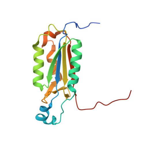
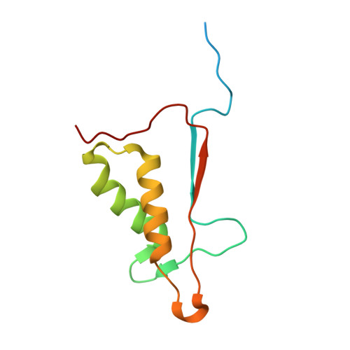
    2C1E, 2C2K, 2C2M, 2C2O, 2C2Z

  • PubMed Abstract: 

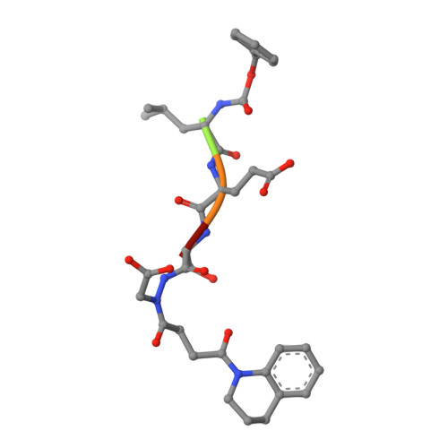
    Aza-peptide Michael acceptors are a novel class of inhibitors that are potent and specific for caspases-2, -3, -6, -7, -8, -9, and -10. The second-order rate constants are in the order of 10(6) M(-1) s(-1). The aza-peptide Michael acceptor inhibitor 18t (Cbz-Asp-Glu-Val-AAsp-trans-CH=CH-CON(CH(2)-1-Naphth)(2) is the most potent compound and it inhibits caspase-3 with a k(2) value of 5620000 M(-1) s(-1). The inhibitor 18t is 13700, 190, 6.4, 594, 37500, and 173-fold more selective for caspase-3 over caspases-2, -6, -7, -8, -9, and -10, respectively. Aza-peptide Michael acceptors designed with caspase specific sequences are selective and do not show any cross reactivity with clan CA cysteine proteases such as papain, cathepsin B, and calpains. High-resolution crystal structures of caspase-3 and caspase-8 in complex with aza-peptide Michael acceptor inhibitors demonstrate the nucleophilic attack on C2 and provide insight into the selectivity and potency of the inhibitors with respect to the P1' moiety.


  • Organizational Affiliation
    • School of Chemistry and Biochemistry, Georgia Institute of Technology, Atlanta, Georgia 30332-0400, USA.

Macromolecule Content 

  • Total Structure Weight: 31.22 kDa 
  • Atom Count: 2,300 
  • Modeled Residue Count: 245 
  • Deposited Residue Count: 270 
  • Unique protein chains: 3

Macromolecules

Find similar proteins by:|  3D Structure
Entity ID: 1
MoleculeChains  Sequence LengthOrganismDetailsImage
CASPASE-8 P18 SUBUNIT159Homo sapiensMutation(s): 0 
EC: 3.4.22 (PDB Primary Data), 3.4.22.61 (UniProt)
UniProt & NIH Common Fund Data Resources
Find proteins for Q14790 (Homo sapiens)
Explore Q14790 
Go to UniProtKB:  Q14790
PHAROS:  Q14790
GTEx:  ENSG00000064012 
Entity Groups
Sequence Clusters30% Identity50% Identity70% Identity90% Identity95% Identity100% Identity
UniProt GroupQ14790
Sequence Annotations
Expand
Reference Sequence
Find similar proteins by:|  3D Structure
Entity ID: 2
MoleculeChains  Sequence LengthOrganismDetailsImage
CASPASE-8 P10 SUBUNIT106Homo sapiensMutation(s): 0 
EC: 3.4.22 (PDB Primary Data), 3.4.22.61 (UniProt)
UniProt & NIH Common Fund Data Resources
Find proteins for Q14790 (Homo sapiens)
Explore Q14790 
Go to UniProtKB:  Q14790
PHAROS:  Q14790
GTEx:  ENSG00000064012 
Entity Groups
Sequence Clusters30% Identity50% Identity70% Identity90% Identity95% Identity100% Identity
UniProt GroupQ14790
Sequence Annotations
Expand
Reference Sequence
Find similar proteins by:  Sequence   |   3D Structure  
Entity ID: 3
MoleculeChains  Sequence LengthOrganismDetailsImage
AZA-PEPTIDE INHIBITOR (5S, 8R, 11S)-8-(2-CARBOXYETHYL) -14-[4-(3,4-DIHYDROQUINOLIN-1(2H)-YL)-4-OXOBUTANOYL] -11-[(1R)-1-HYDROXYETHYL]-5-(2-METHYLPROPYL)-3,6,9,12-TETRAOXO -1-PHENYL-2-OXA-4,7,10,13,14-PENTAAZAHEXADECAN-16-OIC ACID5synthetic constructMutation(s): 0 

Small Molecules

Ligands 1 Unique
IDChains Name / Formula / InChI Key2D Diagram3D Interactions
DTD

Query on DTD


Download:Ideal Coordinates CCD File
D [auth B]DITHIANE DIOL
C4 H8 O2 S2
YPGMOWHXEQDBBV-IMJSIDKUSA-N

Biologically Interesting Molecules (External Reference) 

1 Unique

Experimental Data & Validation

Experimental Data

  • Method: X-RAY DIFFRACTION
  • Resolution: 1.95 Å
  • R-Value Free:  0.187 (Depositor), 0.189 (DCC) 
  • R-Value Work:  0.171 (Depositor), 0.167 (DCC) 
  • R-Value Observed: 0.171 (Depositor) 
Space Group: P 31 2 1
Unit Cell:
Length ( Å )Angle ( ˚ )
a = 62.52α = 90
b = 62.52β = 90
c = 129.94γ = 120
Software Package:
Software NamePurpose
CNSrefinement
XDSdata reduction
XSCALEdata scaling

Structure Validation

View Full Validation Report


Ligand Structure Quality Assessment 


Entry History 

Revision History  (Full details and data files)

  • Version 1.0: 2006-09-20
    Type: Initial release
  • Version 1.1: 2011-07-13
    Changes: Atomic model, Database references, Derived calculations, Non-polymer description, Structure summary, Version format compliance
  • Version 1.2: 2011-07-20
    Changes: Other, Structure summary
  • Version 1.3: 2012-11-30
    Changes: Other
  • Version 1.4: 2017-02-08
    Changes: Source and taxonomy
  • Version 1.5: 2025-04-09
    Changes: Data collection, Database references, Derived calculations, Other, Structure summary