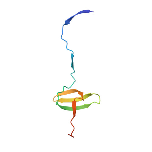
2BZY

Homodimer of CrkL-SH3C domain


Experimental Data Snapshot

  • Method: X-RAY DIFFRACTION
  • Resolution: 2.50 Å
  • R-Value Free: 0.309 
  • R-Value Work: 0.263 
  • R-Value Observed: 0.263 

Starting Model: experimental
View more details

wwPDB Validation   3D Report Full Report


This is version 1.2 of the entry. See complete history


Literature

The C-Terminal SH3 Domain of Crkl as a Dynamic Dimerization Module Transiently Exposing a Nuclear Export Signal.

Harkiolaki, M.Gilbert, R.J.Jones, E.Y.Feller, S.M.

(2006) Structure 14: 1741

  • DOI: https://doi.org/10.1016/j.str.2006.09.013
  • Primary Citation of Related Structures:  
    2BZX, 2BZY

  • PubMed Abstract: 

    CRKL plays essential roles in cell signaling. It consists of an N-terminal SH2 domain followed by two SH3 domains. SH2 and SH3N bind to signaling proteins, but the function of the SH3C domain has remained largely enigmatic. We show here that the SH3C of CRKL forms homodimers in protein crystals and in solution. Evidence for dimer formation of full-length CRKL is also presented. In the SH3C dimer, a nuclear export signal (NES) is mostly buried under the domain surface. The same is true for a monomeric SH3C obtained under different crystallization conditions. Interestingly, partial SH3 unfolding, such as occurs upon dimer/monomer transition, produces a fully-accessible NES through translocation of a single beta strand. Our results document the existence of an SH3 domain dimer formed through exchange of the first SH3 domain beta strand and suggest that partial unfolding of the SH3C is important for the relay of information in vivo.


  • Organizational Affiliation

    Cancer Research UK Cell Signalling Group, Weatherall Institute of Molecular Medicine, University of Oxford, Oxford OX3 9DS, United Kingdom.


Macromolecules
Find similar proteins by:  (by identity cutoff)  |  3D Structure
Entity ID: 1
MoleculeChains Sequence LengthOrganismDetailsImage
CRK-LIKE PROTEIN
A, B
67Homo sapiensMutation(s): 0 
UniProt & NIH Common Fund Data Resources
Find proteins for P46109 (Homo sapiens)
Explore P46109 
Go to UniProtKB:  P46109
PHAROS:  P46109
GTEx:  ENSG00000099942 
Entity Groups  
Sequence Clusters30% Identity50% Identity70% Identity90% Identity95% Identity100% Identity
UniProt GroupP46109
Sequence Annotations
Expand
  • Reference Sequence
Experimental Data & Validation

Experimental Data

  • Method: X-RAY DIFFRACTION
  • Resolution: 2.50 Å
  • R-Value Free: 0.309 
  • R-Value Work: 0.263 
  • R-Value Observed: 0.263 
  • Space Group: P 61 2 2
Unit Cell:
Length ( Å )Angle ( ˚ )
a = 71.317α = 90
b = 71.317β = 90
c = 146.428γ = 120
Software Package:
Software NamePurpose
CNSrefinement
DENZOdata reduction
SCALEPACKdata scaling
AMoREphasing

Structure Validation

View Full Validation Report


Entry History 

Deposition Data

Revision History  (Full details and data files)

  • Version 1.0: 2006-09-28
    Type: Initial release
  • Version 1.1: 2019-05-22
    Changes: Data collection, Refinement description
  • Version 1.2: 2023-12-13
    Changes: Data collection, Database references, Other, Refinement description