2BZD

Galactose recognition by the carbohydrate-binding module of a bacterial sialidase.


Experimental Data Snapshot

  • Method: X-RAY DIFFRACTION
  • Resolution: 2.00 Å
  • R-Value Free: 0.267 
  • R-Value Work: 0.193 
  • R-Value Observed: 0.197 

wwPDB Validation   3D Report Full Report


Ligand Structure Quality Assessment 


This is version 1.3 of the entry. See complete history


Literature

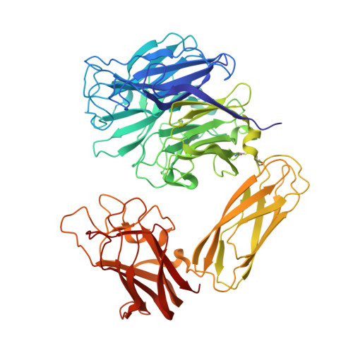
Galactose Recognition by the Carbohydrate-Binding Module of a Bacterial Sialidase.

Newstead, S.L.Watson, J.N.Bennet, A.J.Taylor, G.

(2005) Acta Crystallogr D Biol Crystallogr 61: 1483

  • DOI: https://doi.org/10.1107/S0907444905026132
  • Primary Citation of Related Structures:  
    2BZD

  • PubMed Abstract: 

    Glycoside hydrolases often possess carbohydrate-binding modules (CBMs) in addition to their catalytic domains, which help target the enzymes to appropriate substrates and thereby increase their catalytic efficiency. Sialidases hydrolyse the release of sialic acid from a variety of glycoconjugates and play significant roles in the pathogenesis of a number of important diseases. The sialidase from Micromonospora viridifaciens has a CBM which recognizes galactose. The CBM is linked to the catalytic domain by an immunoglobulin-like domain, resulting in the galactose binding site sitting above the catalytic site, suggesting an interplay between the two sites. By studying nine crystallographically independent structures of the M. viridifaciens sialidase, the relative flexibility of the three domains was analysed. A detailed study is also presented of the recognition of galactose and lactose by the M. viridifaciens CBM. The striking structure of this sialidase suggests a role for the CBM in binding to galactose residues unmasked by the adjacent catalytic site.


  • Organizational Affiliation

    Centre for Biomolecular Sciences, University of St Andrews, St Andrews, Fife KY16 9ST, Scotland.


Macromolecules
Find similar proteins by:  (by identity cutoff)  |  3D Structure
Entity ID: 1
MoleculeChains Sequence LengthOrganismDetailsImage
BACTERIAL SIALIDASE
A, B, C
601Micromonospora viridifaciensMutation(s): 1 
EC: 3.2.1.18
UniProt
Find proteins for Q02834 (Micromonospora viridifaciens)
Explore Q02834 
Go to UniProtKB:  Q02834
Entity Groups  
Sequence Clusters30% Identity50% Identity70% Identity90% Identity95% Identity100% Identity
UniProt GroupQ02834
Sequence Annotations
Expand
  • Reference Sequence
Small Molecules
Ligands 3 Unique
IDChains Name / Formula / InChI Key2D Diagram3D Interactions
GAL
Query on GAL

Download Ideal Coordinates CCD File 
E [auth A],
H [auth B],
K [auth C]
beta-D-galactopyranose
C6 H12 O6
WQZGKKKJIJFFOK-FPRJBGLDSA-N
GOL
Query on GOL

Download Ideal Coordinates CCD File 
F [auth A],
I [auth B],
L [auth C]
GLYCEROL
C3 H8 O3
PEDCQBHIVMGVHV-UHFFFAOYSA-N
NA
Query on NA

Download Ideal Coordinates CCD File 
D [auth A],
G [auth B],
J [auth C]
SODIUM ION
Na
FKNQFGJONOIPTF-UHFFFAOYSA-N
Binding Affinity Annotations 
IDSourceBinding Affinity
GAL Binding MOAD:  2BZD Kd: 1.00e+6 (nM) from 1 assay(s)
Experimental Data & Validation

Experimental Data

  • Method: X-RAY DIFFRACTION
  • Resolution: 2.00 Å
  • R-Value Free: 0.267 
  • R-Value Work: 0.193 
  • R-Value Observed: 0.197 
  • Space Group: P 32 2 1
Unit Cell:
Length ( Å )Angle ( ˚ )
a = 141.457α = 90
b = 141.457β = 90
c = 158.88γ = 120
Software Package:
Software NamePurpose
REFMACrefinement
MOSFLMdata reduction
SCALAdata scaling
AMoREphasing

Structure Validation

View Full Validation Report


Ligand Structure Quality Assessment 


Entry History 

Deposition Data

Revision History  (Full details and data files)

  • Version 1.0: 2005-08-19
    Type: Initial release
  • Version 1.1: 2011-07-13
    Changes: Advisory, Version format compliance
  • Version 1.2: 2020-07-29
    Type: Remediation
    Reason: Carbohydrate remediation
    Changes: Data collection, Derived calculations, Other, Structure summary
  • Version 1.3: 2023-12-13
    Changes: Data collection, Database references, Refinement description, Structure summary