2BXV

Dual binding mode of a novel series of DHODH inhibitors


Experimental Data Snapshot

  • Method: X-RAY DIFFRACTION
  • Resolution: 2.15 Å
  • R-Value Free: 0.223 
  • R-Value Work: 0.203 
  • R-Value Observed: 0.203 

wwPDB Validation   3D Report Full Report


This is version 1.5 of the entry. See complete history


Literature

Dual binding mode of a novel series of DHODH inhibitors.

Baumgartner, R.Walloschek, M.Kralik, M.Gotschlich, A.Tasler, S.Mies, J.Leban, J.

(2006) J Med Chem 49: 1239-1247

  • DOI: https://doi.org/10.1021/jm0506975
  • Primary Citation of Related Structures:  
    2BXV, 2FPT, 2FPV, 2FPY, 2FQI

  • PubMed Abstract: 

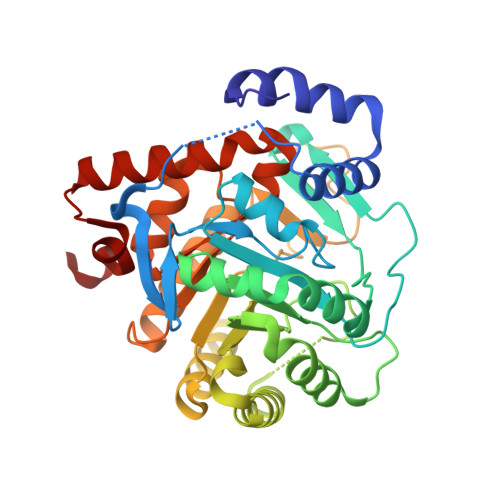
    Human dihydroorotate dehydrogenase (DHODH) represents an important target for the treatment of hyperproliferative and inflammatory diseases. In the cell DHODH catalyzes the rate-limiting step of the de novo pyrimidine biosynthesis. DHODH inhibition results in beneficial immunosuppressant and antiproliferative effects in diseases such as rheumatoid arthritis. Here, we present high-resolution X-ray structures of human DHODH in complex with a novel class of low molecular weight compounds that inhibit the enzyme in the nanomolar range. Some compounds showed an interesting dual binding mode within the same cocrystal strongly depending on the nature of chemical substitution. Measured in vitro activity data correlated with the prevailing mode of binding and explained the observed structure-activity relationship. Additionally, the X-ray data confirmed the competitive nature of the inhibitors toward the putative ubiquinone binding site and will guide structure-based design and synthesis of molecules with higher activity.


  • Organizational Affiliation

    4SC AG, Am Klopferspitz 19a, 82152 Martinsried, Germany. baumgartner@4sc.com


Macromolecules
Find similar proteins by:  (by identity cutoff)  |  3D Structure
Entity ID: 1
MoleculeChains Sequence LengthOrganismDetailsImage
DIHYDROOROTATE DEHYDROGENASE367Homo sapiensMutation(s): 0 
EC: 1.3.3.1
UniProt & NIH Common Fund Data Resources
Find proteins for Q02127 (Homo sapiens)
Explore Q02127 
Go to UniProtKB:  Q02127
PHAROS:  Q02127
GTEx:  ENSG00000102967 
Entity Groups  
Sequence Clusters30% Identity50% Identity70% Identity90% Identity95% Identity100% Identity
UniProt GroupQ02127
Sequence Annotations
Expand
  • Reference Sequence
Small Molecules
Ligands 5 Unique
IDChains Name / Formula / InChI Key2D Diagram3D Interactions
FMN
Query on FMN

Download Ideal Coordinates CCD File 
D [auth A]FLAVIN MONONUCLEOTIDE
C17 H21 N4 O9 P
FVTCRASFADXXNN-SCRDCRAPSA-N
3FT
Query on 3FT

Download Ideal Coordinates CCD File 
F [auth A]2-({[3-FLUORO-3'-(TRIFLUOROMETHOXY)BIPHENYL-4-YL]AMINO}CARBONYL)CYCLOPENT-1-ENE-1-CARBOXYLIC ACID
C20 H15 F4 N O4
CTKBCQCRHRCICI-UHFFFAOYSA-N
ORO
Query on ORO

Download Ideal Coordinates CCD File 
E [auth A]OROTIC ACID
C5 H4 N2 O4
PXQPEWDEAKTCGB-UHFFFAOYSA-N
SO4
Query on SO4

Download Ideal Coordinates CCD File 
B [auth A]SULFATE ION
O4 S
QAOWNCQODCNURD-UHFFFAOYSA-L
ACT
Query on ACT

Download Ideal Coordinates CCD File 
C [auth A]ACETATE ION
C2 H3 O2
QTBSBXVTEAMEQO-UHFFFAOYSA-M
Binding Affinity Annotations 
IDSourceBinding Affinity
3FT BindingDB:  2BXV IC50: 280 (nM) from 1 assay(s)
Binding MOAD:  2BXV IC50: 280 (nM) from 1 assay(s)
Experimental Data & Validation

Experimental Data

  • Method: X-RAY DIFFRACTION
  • Resolution: 2.15 Å
  • R-Value Free: 0.223 
  • R-Value Work: 0.203 
  • R-Value Observed: 0.203 
  • Space Group: P 32 2 1
Unit Cell:
Length ( Å )Angle ( ˚ )
a = 90.652α = 90
b = 90.652β = 90
c = 123.213γ = 120
Software Package:
Software NamePurpose
CNSrefinement
MOSFLMdata reduction
CCP4data scaling
MOLREPphasing

Structure Validation

View Full Validation Report


Entry History 

Deposition Data

Revision History  (Full details and data files)

  • Version 1.0: 2006-08-16
    Type: Initial release
  • Version 1.1: 2011-05-08
    Changes: Version format compliance
  • Version 1.2: 2011-07-13
    Changes: Version format compliance
  • Version 1.3: 2018-04-25
    Changes: Data collection, Database references, Source and taxonomy
  • Version 1.4: 2019-03-06
    Changes: Data collection, Experimental preparation
  • Version 1.5: 2023-12-13
    Changes: Data collection, Database references, Refinement description