2BW7 | pdb_00002bw7

A novel mechanism for adenylyl cyclase inhibition from the crystal structure of its complex with catechol estrogen


Experimental Data Snapshot

  • Method: X-RAY DIFFRACTION
  • Resolution: 2.30 Å
  • R-Value Free: 
    0.257 (Depositor), 0.272 (DCC) 
  • R-Value Work: 
    0.207 (Depositor), 0.227 (DCC) 
  • R-Value Observed: 
    0.207 (Depositor) 

wwPDB Validation 3D Report Full Report

Validation slider image for 2BW7

Ligand Structure Quality Assessment 


This is version 1.3 of the entry. See complete history. 

Literature

A Novel Mechanism for Adenylyl Cyclase Inhibition from the Crystal Structure of its Complex with Catechol Estrogen

Steegborn, C., Litvin, T.N., Hess, K.C., Capper, A.B., Taussig, R., Buck, J., Levin, L.R., Wu, H.

(2005) J Biol Chem 280: 31754

  • DOI: https://doi.org/10.1074/jbc.M507144200
  • Primary Citation Related Structures: 
    2BW7

  • PubMed Abstract: 

    Catechol estrogens are steroid metabolites that elicit physiological responses through binding to a variety of cellular targets. We show here that catechol estrogens directly inhibit soluble adenylyl cyclases and the abundant trans-membrane adenylyl cyclases. Catechol estrogen inhibition is non-competitive with respect to the substrate ATP, and we solved the crystal structure of a catechol estrogen bound to a soluble adenylyl cyclase from Spirulina platensis in complex with a substrate analog. The catechol estrogen is bound to a newly identified, conserved hydrophobic patch near the active center but distinct from the ATP-binding cleft. Inhibitor binding leads to a chelating interaction between the catechol estrogen hydroxyl groups and the catalytic magnesium ion, distorting the active site and trapping the enzyme substrate complex in a non-productive conformation. This novel inhibition mechanism likely applies to other adenylyl cyclase inhibitors, and the identified ligand-binding site has important implications for the development of specific adenylyl cyclase inhibitors.


  • Organizational Affiliation: 
    • Department of Biochemistry, Weill Medical College of Cornell University, New York, New York 10021, USA.

Macromolecule Content 

  • Total Structure Weight: 99.61 kDa 
  • Atom Count: 6,326 
  • Modeled Residue Count: 780 
  • Deposited Residue Count: 876 
  • Unique protein chains: 1

Macromolecules

Find similar proteins by:|  3D Structure
Entity ID: 1
MoleculeChains  Sequence LengthOrganismDetailsImage
ADENYLATE CYCLASE
A, B, C, D
219Limnospira platensisMutation(s): 0 
EC: 4.6.1.1 (PDB Primary Data), 2.7.13.3 (UniProt)
UniProt
Find proteins for O32393 (Limnospira platensis)
Explore O32393 
Go to UniProtKB:  O32393
Entity Groups
Sequence Clusters30% Identity50% Identity70% Identity90% Identity95% Identity100% Identity
UniProt GroupO32393
Sequence Annotations
Expand
Reference Sequence

Small Molecules

Ligands 4 Unique
IDChains Name / Formula / InChI Key2D Diagram3D Interactions
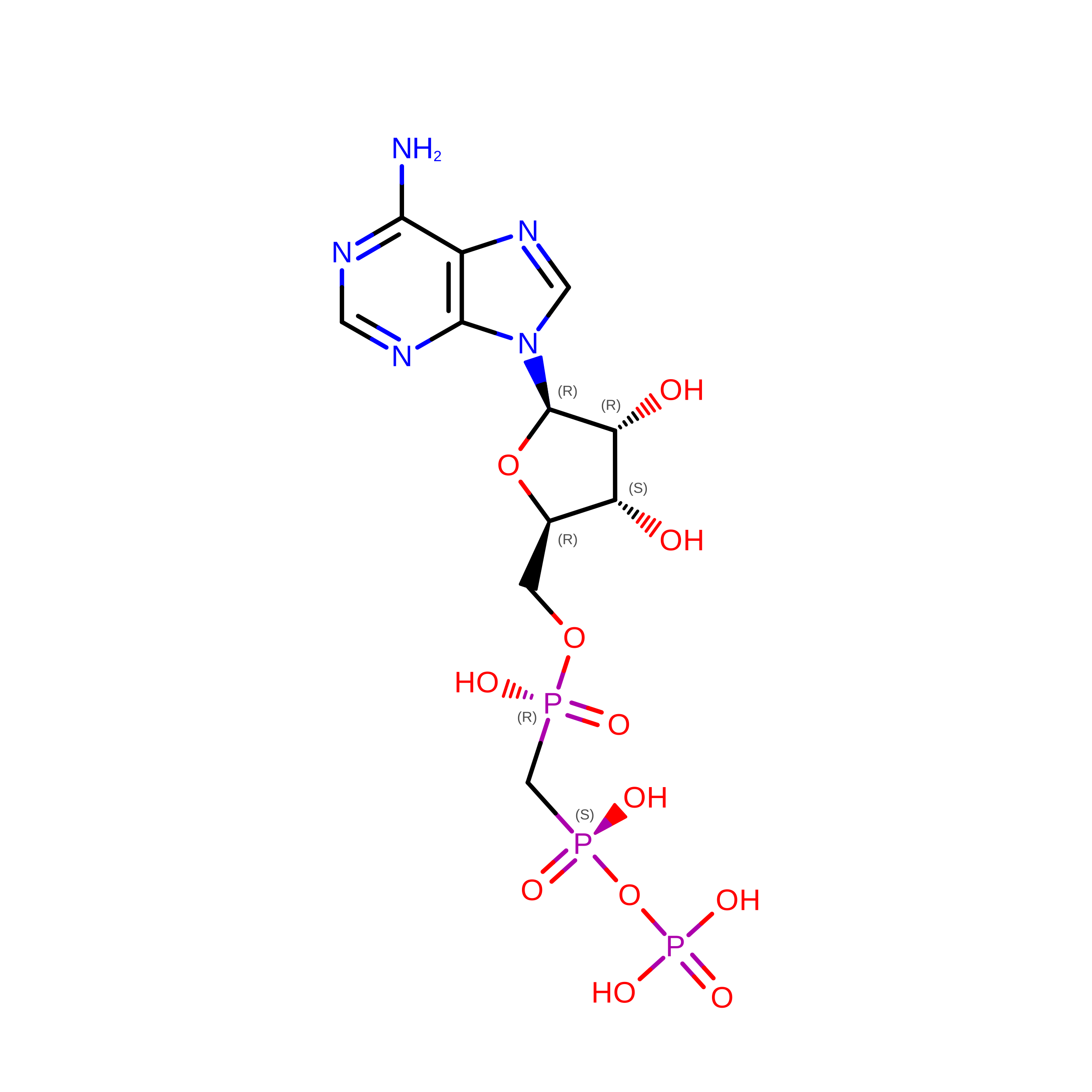
APC

Query on APC


Download:Ideal Coordinates CCD File
E [auth A],
I [auth B],
M [auth C],
P [auth D]
DIPHOSPHOMETHYLPHOSPHONIC ACID ADENOSYL ESTER
C11 H18 N5 O12 P3
CAWZRIXWFRFUQB-IOSLPCCCSA-N
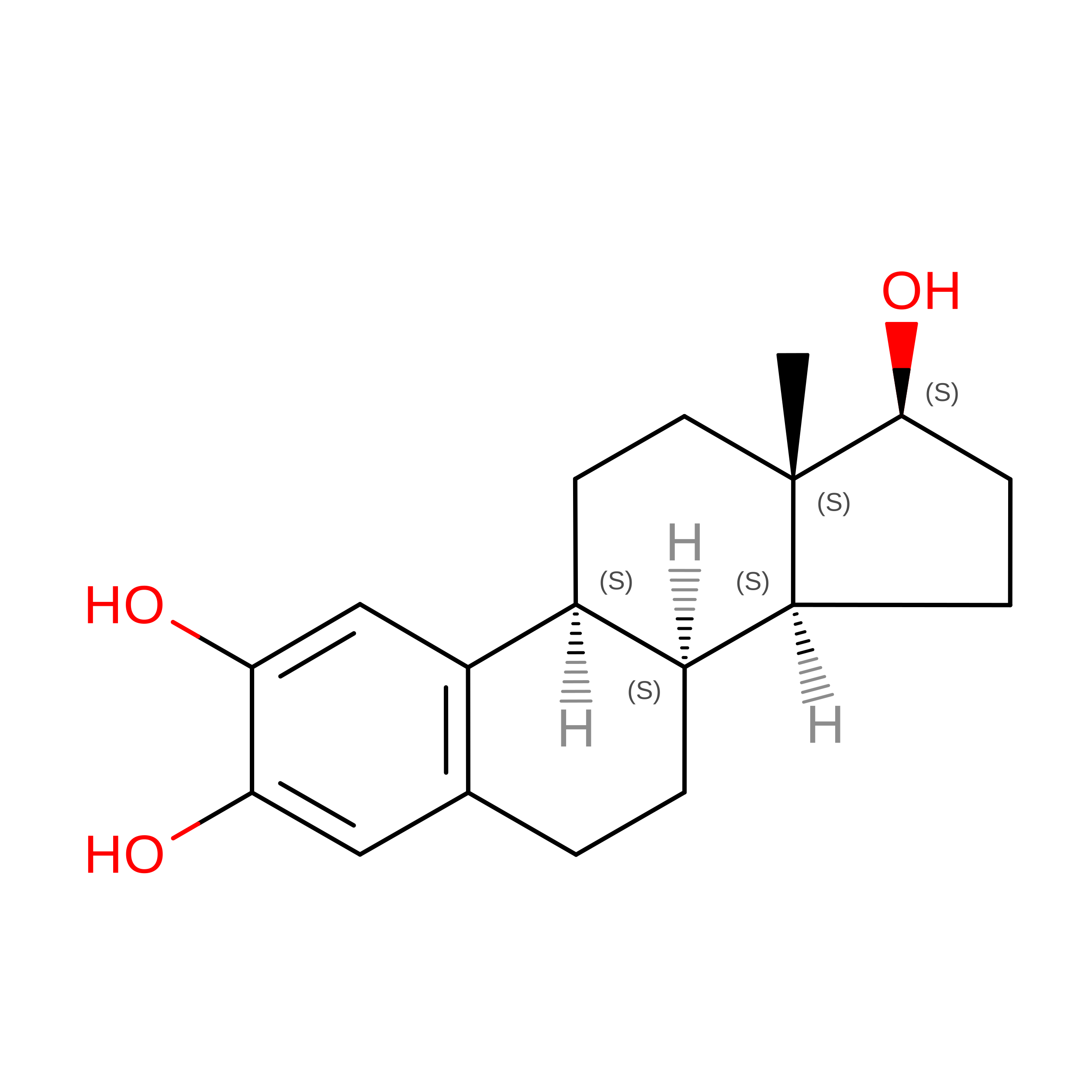
ECS

Query on ECS


Download:Ideal Coordinates CCD File
H [auth A],
L [auth B]
2,3,17BETA-TRIHYDROXY-1,3,5(10)-ESTRATRIENE
C18 H24 O3
DILDHNKDVHLEQB-XSSYPUMDSA-N
CA

Query on CA


Download:Ideal Coordinates CCD File
G [auth A],
K [auth B],
O [auth C],
R [auth D]
CALCIUM ION
Ca
BHPQYMZQTOCNFJ-UHFFFAOYSA-N
MG

Query on MG


Download:Ideal Coordinates CCD File
F [auth A],
J [auth B],
N [auth C],
Q [auth D]
MAGNESIUM ION
Mg
JLVVSXFLKOJNIY-UHFFFAOYSA-N

Experimental Data & Validation

Experimental Data

  • Method: X-RAY DIFFRACTION
  • Resolution: 2.30 Å
  • R-Value Free:  0.257 (Depositor), 0.272 (DCC) 
  • R-Value Work:  0.207 (Depositor), 0.227 (DCC) 
  • R-Value Observed: 0.207 (Depositor) 
Space Group: P 1 21 1
Unit Cell:
Length ( Å )Angle ( ˚ )
a = 53.4α = 90
b = 70.2β = 96
c = 106.7γ = 90
Software Package:
Software NamePurpose
CNSrefinement
DENZOdata reduction
SCALEPACKdata scaling

Structure Validation

View Full Validation Report


Ligand Structure Quality Assessment 


Entry History 

Deposition Data

Revision History  (Full details and data files)

  • Version 1.0: 2005-07-20
    Type: Initial release
  • Version 1.1: 2011-05-08
    Changes: Version format compliance
  • Version 1.2: 2011-07-13
    Changes: Version format compliance
  • Version 1.3: 2024-05-08
    Changes: Data collection, Database references, Derived calculations, Other