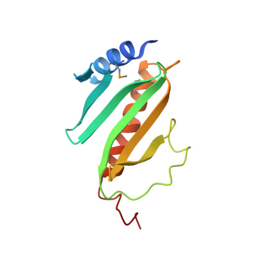
Crystal structure of Yersinia enterocolitica type III secretion chaperone SycT.
Buttner, C.R., Cornelis, G.R., Heinz, D.W., Niemann, H.H.(2005) Protein Sci 14: 1993-2002
- PubMed: 16046625 Search on PubMedSearch on PubMed Central
- DOI: https://doi.org/10.1110/ps.051474605
- Primary Citation Related Structures:
2BSH, 2BSI, 2BSJ - PubMed Abstract:
Pathogenic Yersinia species use a type III secretion (TTS) system to deliver a number of cytotoxic effector proteins directly into the mammalian host cell. To ensure effective translocation, several such effector proteins transiently bind to specific chaperones in the bacterial cytoplasm. Correspondingly, SycT is the chaperone of YopT, a cysteine protease that cleaves the membrane-anchor of Rho-GTPases in the host. We have analyzed the complex between YopT and SycT and determined the structure of SycT in three crystal forms. Biochemical studies indicate a stoichometric effector/chaperone ratio of 1:2 and the chaperone-binding site contains at least residues 52-103 of YopT. The crystal structures reveal a SycT homodimer with an overall fold similar to that of other TTS effector chaperones. In contrast to the canonical five-stranded anti-parallel beta-sheet flanked by three alpha-helices, SycT lacks the dimerization alpha-helix and has an additional beta-strand capable of undergoing a conformational change. The dimer interface consists of two beta-strands and the connecting loops. Two hydrophobic patches involved in effector binding in other TTS effector chaperones are also found in SycT. The structural similarity of SycT to other chaperones and the spatial conservation of effector-binding sites support the idea that TTS effector chaperones form a single functional and structural group.
- Division of Structural Biology, German Research Centre for Biotechnology, D-38124, Braunschweig, Germany.
Organizational Affiliation: