Structure of an Atypical Epoxide Hydrolase from Mycobacterium Tuberculosis Gives Insights Into its Function.
Johansson, P., Unge, T., Cronin, A., Arand, M., Bergfors, T., Jones, T.A., Mowbray, S.L.(2005) J Mol Biology 351: 1048
- PubMed: 16051262 Search on PubMed
- DOI: https://doi.org/10.1016/j.jmb.2005.06.055
- Primary Citation Related Structures:
2BNG - PubMed Abstract:
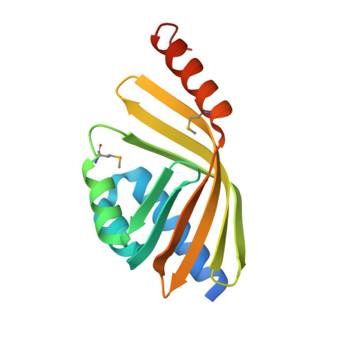
Epoxide hydrolases are vital to many organisms by virtue of their roles in detoxification, metabolism and processing of signaling molecules. The Mycobacterium tuberculosis genome encodes an unusually large number of epoxide hydrolases, suggesting that they might be of particular importance to these bacteria. We report here the first structure of an epoxide hydrolase from M.tuberculosis, solved to a resolution of 2.5 A using single-wavelength anomalous dispersion (SAD) from a selenomethionine-substituted protein. The enzyme features a deep active-site pocket created by the packing of three helices onto a curved six-stranded beta-sheet. This structure is similar to a previously described limonene-1,2-epoxide hydrolase from Rhodococcus erythropolis and unlike the alpha/beta-hydrolase fold typical of mammalian epoxide hydrolases (EH). A number of changes in the mycobacterial enzyme create a wider and deeper substrate-binding pocket than is found in its Rhodococcus homologue. Interestingly, each structure contains a different type of endogenous ligand of unknown origin bound in its active site. As a consequence of its wider substrate-binding pocket, the mycobacterial EH is capable of hydrolyzing long or bulky lipophilic epoxides such as 10,11-epoxystearic acid and cholesterol 5,6-oxide at appreciable rates, suggesting that similar compound(s) will serve as its physiological substrate(s).
- Department of Cell and Molecular Biology, Uppsala University, Biomedical Center, Box 596, SE-751 24 Uppsala, Sweden.
Organizational Affiliation: