2BMG

Crystal structure of factor Xa in complex with 50


Experimental Data Snapshot

  • Method: X-RAY DIFFRACTION
  • Resolution: 2.70 Å
  • R-Value Free: 0.277 
  • R-Value Work: 0.193 

Starting Model: experimental
View more details

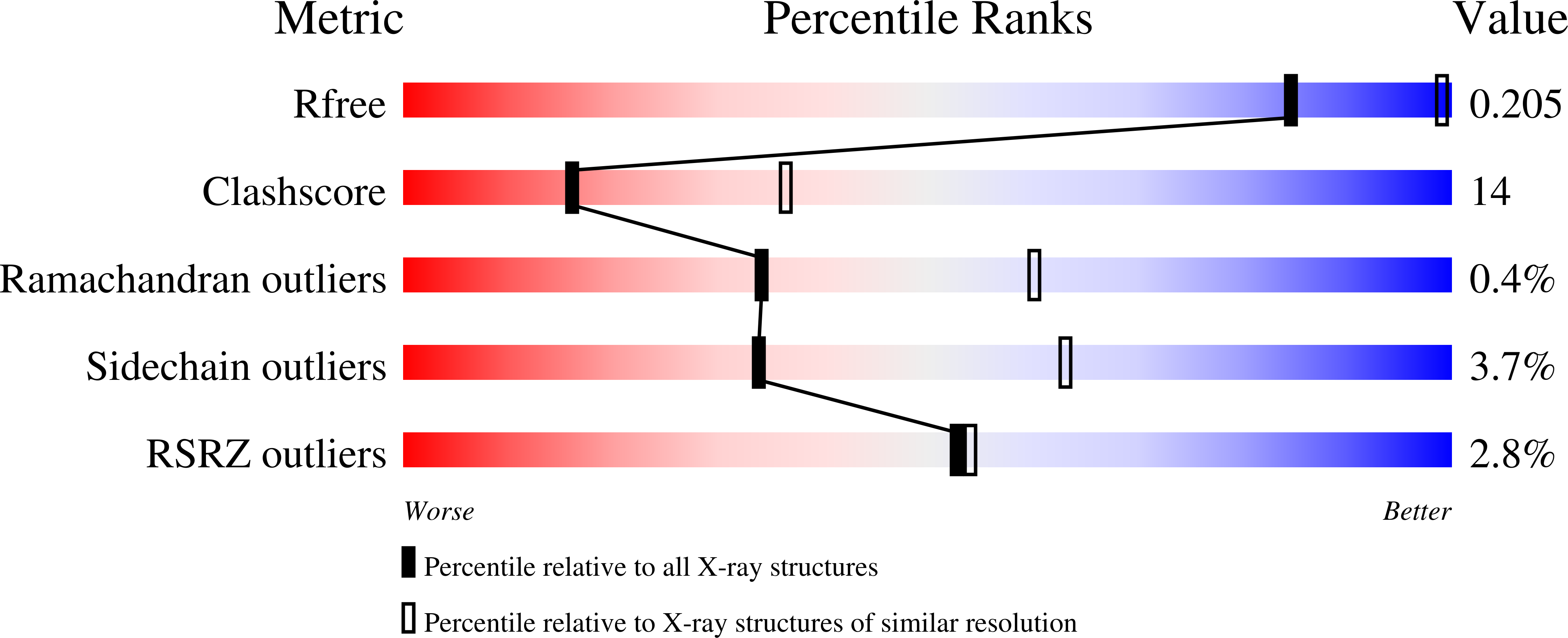
wwPDB Validation   3D Report Full Report


Ligand Structure Quality Assessment 


This is version 1.5 of the entry. See complete history


Literature

Structural Requirements for Factor Xa Inhibition by 3-Oxybenzamides with Neutral P1 Substituents: Combining X-Ray Crystallography, 3D-Qsar and Tailored Scoring Functions

Matter, H.Will, D.W.Nazare, M.Schreuder, H.Laux, V.Wehner, V.Liesum, A.

(2005) J Med Chem 48: 3290

  • DOI: https://doi.org/10.1021/jm049187l
  • Primary Citation of Related Structures:  
    2BMG

  • PubMed Abstract: 

    The design, synthesis, and structure-activity relationship of 3-oxybenzamides as potent inhibitors of the coagulation protease factor Xa are described on the basis of X-ray structures, privileged structure motifs, and SAR information. A total of six X-ray structures of fXa/inhibitor complexes led us to identify the major protein-ligand interactions. The binding mode is characterized by a lipophilic dichlorophenyl substituent interacting with Tyr228 in the protease S1 pocket, while polar parts are accommodated in S4. This alignment in combination with docking allowed derivation of 3D-QSAR models and tailored scoring functions to rationalize biological affinity and provide guidelines for optimization. The resulting models showed good correlation coefficients and predictions of external test sets. Furthermore, they correspond to binding site topologies in terms of steric, electrostatic, and hydrophobic complementarity. Two approaches to derive tailored scoring functions combining binding site and ligand information led to predictive models with acceptable predictions of the external set. Good correlations to experimental affinities were obtained for both AFMoC (adaptation of fields for molecular comparison) and the novel TScore function. The SAR information from 3D-QSAR and tailored scoring functions agrees with all experimental data and provides guidelines and reasonable activity estimations for novel fXa inhibitors.


  • Organizational Affiliation

    DI and A Chemistry, Aventis Pharma Deutschland GmbH, A Company of the Sanofi-Aventis Group, Building G 878, D-65926 Frankfurt am Main, Germany. hans.matter@aventis.com


Macromolecules
Find similar proteins by:  (by identity cutoff)  |  3D Structure
Entity ID: 1
MoleculeChains Sequence LengthOrganismDetailsImage
COAGULATION FACTOR X53Homo sapiensMutation(s): 0 
EC: 3.4.21.6
UniProt & NIH Common Fund Data Resources
Find proteins for P00742 (Homo sapiens)
Explore P00742 
Go to UniProtKB:  P00742
PHAROS:  P00742
GTEx:  ENSG00000126218 
Entity Groups  
Sequence Clusters30% Identity50% Identity70% Identity90% Identity95% Identity100% Identity
UniProt GroupP00742
Sequence Annotations
Expand
  • Reference Sequence
Find similar proteins by:  (by identity cutoff)  |  3D Structure
Entity ID: 2
MoleculeChains Sequence LengthOrganismDetailsImage
COAGULATION FACTOR X234Homo sapiensMutation(s): 0 
EC: 3.4.21.6
UniProt & NIH Common Fund Data Resources
Find proteins for P00742 (Homo sapiens)
Explore P00742 
Go to UniProtKB:  P00742
PHAROS:  P00742
GTEx:  ENSG00000126218 
Entity Groups  
Sequence Clusters30% Identity50% Identity70% Identity90% Identity95% Identity100% Identity
UniProt GroupP00742
Sequence Annotations
Expand
  • Reference Sequence
Small Molecules
Ligands 2 Unique
IDChains Name / Formula / InChI Key2D Diagram3D Interactions
I1H
Query on I1H

Download Ideal Coordinates CCD File 
D [auth B]3-[2-(2,4-DICHLOROPHENYL)ETHOXY]-4-METHOXY-N-[(1-PYRIDIN-4-YLPIPERIDIN-4-YL)METHYL]BENZAMIDE
C27 H29 Cl2 N3 O3
UVSAHJWEJXWVGO-UHFFFAOYSA-N
CA
Query on CA

Download Ideal Coordinates CCD File 
C [auth B]CALCIUM ION
Ca
BHPQYMZQTOCNFJ-UHFFFAOYSA-N
Binding Affinity Annotations 
IDSourceBinding Affinity
I1H PDBBind:  2BMG Ki: 18 (nM) from 1 assay(s)
BindingDB:  2BMG Ki: 18 (nM) from 1 assay(s)
Experimental Data & Validation

Experimental Data

  • Method: X-RAY DIFFRACTION
  • Resolution: 2.70 Å
  • R-Value Free: 0.277 
  • R-Value Work: 0.193 
  • Space Group: P 21 21 21
Unit Cell:
Length ( Å )Angle ( ˚ )
a = 57α = 90
b = 72.3β = 90
c = 78.1γ = 90
Software Package:
Software NamePurpose
CNXrefinement
XDSdata reduction
XSCALEdata scaling
CNXphasing

Structure Validation

View Full Validation Report


Ligand Structure Quality Assessment 


Entry History 

Deposition Data

Revision History  (Full details and data files)

  • Version 1.0: 2006-03-08
    Type: Initial release
  • Version 1.1: 2011-05-08
    Changes: Version format compliance
  • Version 1.2: 2011-07-13
    Changes: Version format compliance
  • Version 1.3: 2017-06-28
    Changes: Data collection
  • Version 1.4: 2019-01-30
    Changes: Data collection, Experimental preparation
  • Version 1.5: 2023-12-13
    Changes: Data collection, Database references, Derived calculations, Other, Refinement description