2BME | pdb_00002bme

high resolution structure of GppNHp-bound human Rab4a


Experimental Data Snapshot

  • Method: X-RAY DIFFRACTION
  • Resolution: 1.57 Å
  • R-Value Free: 
    0.229 (Depositor), 0.233 (DCC) 
  • R-Value Work: 
    0.192 (Depositor), 0.204 (DCC) 
  • R-Value Observed: 
    0.194 (Depositor) 

Starting Model: experimental
View more details

wwPDB Validation 3D Report Full Report

Validation slider image for 2BME

Ligand Structure Quality Assessment 


This is version 1.4 of the entry. See complete history

Literature

High Resolution Crystal Structures of Human Rab4A in its Active and Inactive Conformations.

Huber, S.K.Scheidig, A.J.

(2005) FEBS Lett 579: 2821

  • DOI: https://doi.org/10.1016/j.febslet.2005.04.020
  • Primary Citation Related Structures: 
    2BMD, 2BME

  • PubMed Abstract: 

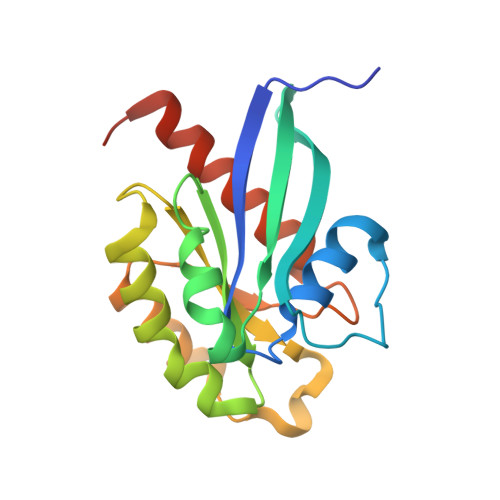
    The Ras-related human GTPase Rab4a is involved in the regulation of endocytosis through the sorting and recycling of early endosomes. Towards further insight, we have determined the three-dimensional crystal structure of human Rab4a in its GppNHp-bound state to 1.6 Angstroms resolution and in its GDP-bound state to 1.8 Angstroms resolution, respectively. Despite the similarity of the overall structure with other Rab proteins, Rab4a displays significant differences. The structures are discussed with respect to the recently determined structure of human Rab5a and its complex with the Rab5-binding domain of the bivalent effector Rabaptin-5. The Rab4 specific residue His39 modulates the nucleotide binding pocket giving rise to a reduced rate for nucleotide hydrolysis and exchange. In comparison to Rab5, Rab4a has a different GDP-bound conformation within switch 1 region and displays shifts in position and orientation of the hydrophobic triad. The observed differences at the S2-L3-S3 region represent a new example of structural plasticity among Rab proteins and may provide a structural basis to understand the differential binding of similar effector proteins.


  • Organizational Affiliation
    • Max-Planck Institut für Molekulare Physiologie, Abteilung für Physikalische Biochemie, Dortmund, Germany.

Macromolecule Content 

  • Total Structure Weight: 86.79 kDa 
  • Atom Count: 6,403 
  • Modeled Residue Count: 697 
  • Deposited Residue Count: 744 
  • Unique protein chains: 1

Macromolecules

Find similar proteins by:|  3D Structure
Entity ID: 1
MoleculeChains  Sequence LengthOrganismDetailsImage
RAS-RELATED PROTEIN RAB4A
A, B, C, D
186Homo sapiensMutation(s): 0 
EC: 3.6.5.2
UniProt & NIH Common Fund Data Resources
Find proteins for P20338 (Homo sapiens)
Explore P20338 
Go to UniProtKB:  P20338
PHAROS:  P20338
GTEx:  ENSG00000168118 
Entity Groups
Sequence Clusters30% Identity50% Identity70% Identity90% Identity95% Identity100% Identity
UniProt GroupP20338
Sequence Annotations
Expand
Reference Sequence

Small Molecules

Ligands 4 Unique
IDChains Name / Formula / InChI Key2D Diagram3D Interactions
GNP

Query on GNP


Download:Ideal Coordinates CCD File
E [auth A],
J [auth B],
L [auth C],
Q [auth D]
PHOSPHOAMINOPHOSPHONIC ACID-GUANYLATE ESTER
C10 H17 N6 O13 P3
UQABYHGXWYXDTK-UUOKFMHZSA-N
TRS

Query on TRS


Download:Ideal Coordinates CCD File
F [auth A],
G [auth A],
M [auth C],
P [auth D]
2-AMINO-2-HYDROXYMETHYL-PROPANE-1,3-DIOL
C4 H12 N O3
LENZDBCJOHFCAS-UHFFFAOYSA-O
BME

Query on BME


Download:Ideal Coordinates CCD File
H [auth A],
N [auth C]
BETA-MERCAPTOETHANOL
C2 H6 O S
DGVVWUTYPXICAM-UHFFFAOYSA-N
MG

Query on MG


Download:Ideal Coordinates CCD File
I [auth A],
K [auth B],
O [auth C],
R [auth D]
MAGNESIUM ION
Mg
JLVVSXFLKOJNIY-UHFFFAOYSA-N

Experimental Data & Validation

Experimental Data

  • Method: X-RAY DIFFRACTION
  • Resolution: 1.57 Å
  • R-Value Free:  0.229 (Depositor), 0.233 (DCC) 
  • R-Value Work:  0.192 (Depositor), 0.204 (DCC) 
  • R-Value Observed: 0.194 (Depositor) 
Space Group: P 43
Unit Cell:
Length ( Å )Angle ( ˚ )
a = 99.79α = 90
b = 99.79β = 90
c = 95.54γ = 90
Software Package:
Software NamePurpose
REFMACrefinement
XDSdata reduction
XSCALEdata scaling
MOLREPphasing

Structure Validation

View Full Validation Report


Ligand Structure Quality Assessment 


Entry History 

Deposition Data

Revision History  (Full details and data files)

  • Version 1.0: 2005-04-25
    Type: Initial release
  • Version 1.1: 2011-05-08
    Changes: Version format compliance
  • Version 1.2: 2011-07-13
    Changes: Version format compliance
  • Version 1.3: 2019-05-08
    Changes: Data collection, Experimental preparation, Other
  • Version 1.4: 2023-12-13
    Changes: Data collection, Database references, Derived calculations, Other, Refinement description