2BM3 | pdb_00002bm3

Structure of the Type II cohesin from Clostridium thermocellum SdbA


Experimental Data Snapshot

  • Method: X-RAY DIFFRACTION
  • Resolution: 1.80 Å
  • R-Value Free: 
    0.260 (Depositor), 0.261 (DCC) 
  • R-Value Work: 
    0.189 (Depositor), 0.188 (DCC) 
  • R-Value Observed: 
    0.192 (Depositor) 

wwPDB Validation 3D Report Full Report

Validation slider image for 2BM3

This is version 1.2 of the entry. See complete history

Literature

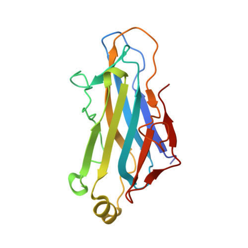
Insights Into the Structural Determinants of Cohesin-Dockerin Specificity Revealed by the Crystal Structure of the Type II Cohesin from Clostridium Thermocellum Sdba.

Carvalho, A.L.Pires, V.M.R.Gloster, T.M.Turkenburg, J.P.Prates, J.A.M.Ferreira, L.M.A.Romao, M.J.Davies, G.J.Fontes, C.M.G.A.Gilbert, H.J.

(2005) J Mol Biology 349: 909

  • DOI: https://doi.org/10.1016/j.jmb.2005.04.037
  • Primary Citation Related Structures: 
    2BM3

  • PubMed Abstract: 

    The plant cell wall degrading enzymes expressed by anaerobic microorganisms form large multienzyme complexes (cellulosomes). Cellulosomes assemble by the Type I dockerins on the catalytic subunits binding to the reiterated Type I cohesins in the molecular scaffold, while Type II dockerin-cohesin interactions anchor the complex onto the bacterial cell surface. Type I and Type II cohesin, dockerin pairs show no cross-specificity. Here we report the crystal structure of the Type II cohesin (CohII) from the Clostridium thermocellum cell surface anchoring protein SdbA. The protein domain contains nine beta-strands and a small alpha-helix. The beta-strands assemble into two elongated beta-sheets that display a typical jelly roll fold. The structure of CohII is very similar to Type I cohesins, and the dockerin binding site, which is centred at beta-strands 3, 5 and 6, is likely to be conserved in the two proteins. Subtle differences in the topology of the binding sites and a lack of sequence identity in the beta-strands that comprise the core of the dockerin binding site explain why Type I and Type II cohesins display such distinct specificities for their target dockerins.


  • Organizational Affiliation
    • REQUIMTE/CQFB, Departamento de Química, Faculdade de Ciências e Tecnologia, Universidade Nova de Lisboa, 2829-516 Caparica, Portugal.

Macromolecule Content 

  • Total Structure Weight: 18.3 kDa 
  • Atom Count: 1,532 
  • Modeled Residue Count: 162 
  • Deposited Residue Count: 166 
  • Unique protein chains: 1

Macromolecules

Find similar proteins by:|  3D Structure
Entity ID: 1
MoleculeChains  Sequence LengthOrganismDetailsImage
SCAFFOLDING DOCKERIN BINDING PROTEIN A166Acetivibrio thermocellusMutation(s): 0 
UniProt
Find proteins for P71143 (Acetivibrio thermocellus)
Explore P71143 
Go to UniProtKB:  P71143
Entity Groups
Sequence Clusters30% Identity50% Identity70% Identity90% Identity95% Identity100% Identity
UniProt GroupP71143
Sequence Annotations
Expand
Reference Sequence

Experimental Data & Validation

Experimental Data

  • Method: X-RAY DIFFRACTION
  • Resolution: 1.80 Å
  • R-Value Free:  0.260 (Depositor), 0.261 (DCC) 
  • R-Value Work:  0.189 (Depositor), 0.188 (DCC) 
  • R-Value Observed: 0.192 (Depositor) 
Space Group: P 63 2 2
Unit Cell:
Length ( Å )Angle ( ˚ )
a = 79.677α = 90
b = 79.677β = 90
c = 101.745γ = 120
Software Package:
Software NamePurpose
DENZOdata reduction
SCALEPACKdata scaling
SHARPphasing
DMphasing
REFMACrefinement

Structure Validation

View Full Validation Report


Entry History 

Revision History  (Full details and data files)

  • Version 1.0: 2005-03-10
    Type: Initial release
  • Version 1.1: 2013-03-06
    Changes: Data collection, Database references, Derived calculations, Non-polymer description, Other, Structure summary, Version format compliance
  • Version 1.2: 2024-05-08
    Changes: Data collection, Database references, Derived calculations, Other