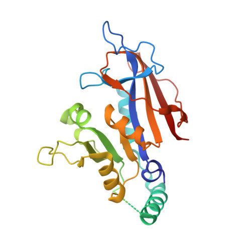
2BLC

SP21 double mutant P. vivax Dihydrofolate reductase in complex with des-chloropyrimethamine


Experimental Data Snapshot

  • Method: X-RAY DIFFRACTION
  • Resolution: 2.25 Å
  • R-Value Free: 0.280 
  • R-Value Work: 0.215 
  • R-Value Observed: 0.215 

wwPDB Validation   3D Report Full Report


This is version 1.4 of the entry. See complete history


Literature

Crystal Structure of Dihydrofolate Reductase from Plasmodium Vivax: Pyrimethamine Displacement Linked with Mutation-Induced Resistance.

Kongsaeree, P.Khongsuk, P.Leartsakulpanich, U.Chitnumsub, P.Tarnchompoo, B.Walkinshaw, M.D.Yuthavong, Y.

(2005) Proc Natl Acad Sci U S A 102: 13046

  • DOI: https://doi.org/10.1073/pnas.0501747102
  • Primary Citation of Related Structures:  
    2BL9, 2BLA, 2BLB, 2BLC

  • PubMed Abstract: 

    Pyrimethamine (Pyr) targets dihydrofolate reductase of Plasmodium vivax (PvDHFR) as well as other malarial parasites, but its use as antimalarial is hampered by the widespread high resistance. Comparison of the crystal structures of PvDHFR from wild-type and the Pyr-resistant (SP21, Ser-58 --> Arg + Ser-117 --> Asn) strain as complexes with NADPH and Pyr or its analog lacking p-Cl (Pyr20) clearly shows that the steric conflict arising from the side chain of Asn-117 in the mutant enzyme, accompanied by the loss of binding to Ser-120, is mainly responsible for the reduction in binding of Pyr. Pyr20 still effectively inhibits both the wild-type and SP21 proteins, and the x-ray structures of these complexes show how Pyr20 fits into both active sites without steric strain. These structural insights suggest a general approach for developing new generations of antimalarial DHFR inhibitors that, by only occupying substrate space of the active site, would retain binding affinity with the mutant enzymes.


  • Organizational Affiliation

    Department of Chemistry and Center for Protein Structure and Function, Faculty of Science, Mahidol University, Rama 6 Road, Bangkok 10400, Thailand.


Macromolecules
Find similar proteins by:  (by identity cutoff)  |  3D Structure
Entity ID: 1
MoleculeChains Sequence LengthOrganismDetailsImage
DIHYDROFOLATE REDUCTASE-THYMIDYLATE SYNTHASE238Plasmodium vivaxMutation(s): 0 
EC: 1.5.1.3 (PDB Primary Data), 2.1.1.45 (UniProt)
UniProt
Find proteins for O02604 (Plasmodium vivax)
Explore O02604 
Go to UniProtKB:  O02604
Entity Groups  
Sequence Clusters30% Identity50% Identity70% Identity90% Identity95% Identity100% Identity
UniProt GroupO02604
Sequence Annotations
Expand
  • Reference Sequence
Small Molecules
Binding Affinity Annotations 
IDSourceBinding Affinity
CP7 BindingDB:  2BLC Ki: min: 0.03, max: 0.08 (nM) from 2 assay(s)
IC50: min: 1520, max: 1820 (nM) from 2 assay(s)
Experimental Data & Validation

Experimental Data

  • Method: X-RAY DIFFRACTION
  • Resolution: 2.25 Å
  • R-Value Free: 0.280 
  • R-Value Work: 0.215 
  • R-Value Observed: 0.215 
  • Space Group: C 1 2 1
Unit Cell:
Length ( Å )Angle ( ˚ )
a = 132α = 90
b = 56.31β = 107.5
c = 45.67γ = 90
Software Package:
Software NamePurpose
CNSrefinement
CrystalCleardata reduction
CrystalCleardata scaling
AMoREphasing

Structure Validation

View Full Validation Report


Entry History 

Deposition Data

Revision History  (Full details and data files)

  • Version 1.0: 2005-09-07
    Type: Initial release
  • Version 1.1: 2011-05-08
    Changes: Version format compliance
  • Version 1.2: 2011-07-13
    Changes: Version format compliance
  • Version 1.3: 2019-05-22
    Changes: Data collection, Other, Refinement description
  • Version 1.4: 2024-05-08
    Changes: Data collection, Database references, Other