2BKE | pdb_00002bke

Conformational Flexibility Revealed by the Crystal Structure of a Crenarchaeal RadA


Experimental Data Snapshot

  • Method: X-RAY DIFFRACTION
  • Resolution: 3.20 Å
  • R-Value Free: 
    0.240 (Depositor), 0.246 (DCC) 
  • R-Value Work: 
    0.174 (Depositor), 0.183 (DCC) 
  • R-Value Observed: 
    0.178 (Depositor) 

Starting Model: experimental
View more details

wwPDB Validation 3D Report Full Report

Validation slider image for 2BKE

This is version 1.4 of the entry. See complete history

Literature

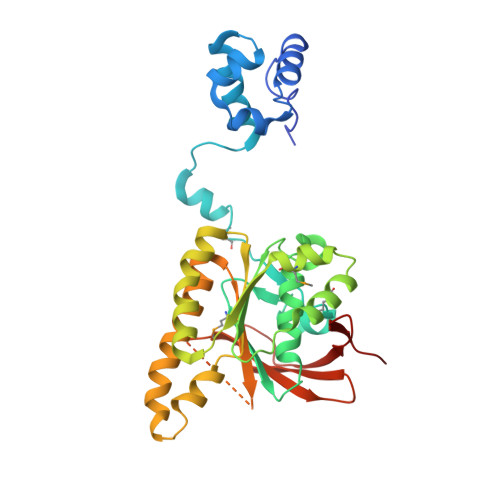
Conformational Flexibility Revealed by the Crystal Structure of a Crenarchaeal Rada

Ariza, A.Richard, D.L.White, M.F.Bond, C.S.

(2005) Nucleic Acids Res 33: 1465

  • DOI: https://doi.org/10.1093/nar/gki288
  • Primary Citation Related Structures: 
    2BKE

  • PubMed Abstract: 

    Homologous recombinational repair is an essential mechanism for repair of double-strand breaks in DNA. Recombinases of the RecA-fold family play a crucial role in this process, forming filaments that utilize ATP to mediate their interactions with single- and double-stranded DNA. The recombinase molecules present in the archaea (RadA) and eukaryota (Rad51) are more closely related to each other than to their bacterial counterpart (RecA) and, as a result, RadA makes a suitable model for the eukaryotic system. The crystal structure of Sulfolobus solfataricus RadA has been solved to a resolution of 3.2 A in the absence of nucleotide analogues or DNA, revealing a narrow filamentous assembly with three molecules per helical turn. As observed in other RecA-family recombinases, each RadA molecule in the filament is linked to its neighbour via interactions of a short beta-strand with the neighbouring ATPase domain. However, despite apparent flexibility between domains, comparison with other structures indicates conservation of a number of key interactions that introduce rigidity to the system, allowing allosteric control of the filament by interaction with ATP. Additional analysis reveals that the interaction specificity of the five human Rad51 paralogues can be predicted using a simple model based on the RadA structure.


  • Organizational Affiliation
    • Division of Biological Chemistry and Molecular Microbiology, School of Life Sciences, University of Dundee Dow St, Dundee, DD1 5EH, UK.

Macromolecule Content 

  • Total Structure Weight: 36.28 kDa 
  • Atom Count: 2,327 
  • Modeled Residue Count: 293 
  • Deposited Residue Count: 324 
  • Unique protein chains: 1

Macromolecules

Find similar proteins by:|  3D Structure
Entity ID: 1
MoleculeChains  Sequence LengthOrganismDetailsImage
DNA REPAIR AND RECOMBINATION PROTEIN RADA324Saccharolobus solfataricus P2Mutation(s): 0 
UniProt
Find proteins for Q55075 (Saccharolobus solfataricus (strain ATCC 35092 / DSM 1617 / JCM 11322 / P2))
Explore Q55075 
Go to UniProtKB:  Q55075
Entity Groups
Sequence Clusters30% Identity50% Identity70% Identity90% Identity95% Identity100% Identity
UniProt GroupQ55075
Sequence Annotations
Expand
Reference Sequence

Small Molecules

Ligands 1 Unique
IDChains Name / Formula / InChI Key2D Diagram3D Interactions
CL

Query on CL


Download:Ideal Coordinates CCD File
B [auth A]CHLORIDE ION
Cl
VEXZGXHMUGYJMC-UHFFFAOYSA-M
Modified Residues  1 Unique
IDChains TypeFormula2D DiagramParent
MSE
Query on MSE
A
L-PEPTIDE LINKINGC5 H11 N O2 SeMET

Experimental Data & Validation

Experimental Data

  • Method: X-RAY DIFFRACTION
  • Resolution: 3.20 Å
  • R-Value Free:  0.240 (Depositor), 0.246 (DCC) 
  • R-Value Work:  0.174 (Depositor), 0.183 (DCC) 
  • R-Value Observed: 0.178 (Depositor) 
Space Group: P 31 2 1
Unit Cell:
Length ( Å )Angle ( ˚ )
a = 98.146α = 90
b = 98.146β = 90
c = 99.19γ = 120
Software Package:
Software NamePurpose
REFMACrefinement
DENZOdata reduction
SCALEPACKdata scaling
MOLREPphasing

Structure Validation

View Full Validation Report


Entry History 

Deposition Data

Revision History  (Full details and data files)

  • Version 1.0: 2005-03-16
    Type: Initial release
  • Version 1.1: 2011-07-13
    Changes: Advisory, Version format compliance
  • Version 1.2: 2018-01-24
    Changes: Source and taxonomy
  • Version 1.3: 2023-12-13
    Changes: Data collection, Database references, Derived calculations, Other, Refinement description
  • Version 1.4: 2024-10-09
    Changes: Structure summary