2BIB | pdb_00002bib

Crystal structure of the complete modular teichioic acid phosphorylcholine esterase Pce (CbpE) from Streptococcus pneumoniae


Experimental Data Snapshot

  • Method: X-RAY DIFFRACTION
  • Resolution: 1.92 Å
  • R-Value Free: 
    0.233 (Depositor) 
  • R-Value Work: 
    0.194 (Depositor) 
  • R-Value Observed: 
    0.197 (Depositor) 

wwPDB Validation 3D Report Full Report

Validation slider image for 2BIB

Ligand Structure Quality Assessment 


This is version 1.3 of the entry. See complete history

Literature

Insights Into Pneumococcal Pathogenesis from Crystal Structure of the Modular Teichoic Acid Phosphorylcholine Esterase Pce

Hermoso, J.A.Lagartera, L.Gonzalez, A.Stelter, M.Garcia, P.Martinez-Ripoll, M.Garcia, J.L.Menendez, M.

(2005) Nat Struct Mol Biol 12: 533

  • DOI: https://doi.org/10.1038/nsmb940
  • Primary Citation Related Structures: 
    2BIB

  • PubMed Abstract: 

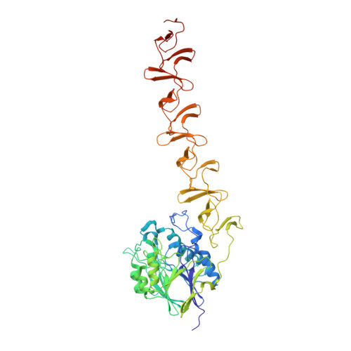
    Phosphorylcholine, a specific component of the pneumococcal cell wall, is crucial in pathogenesis. It directly binds to the human platelet-activating factor (PAF) receptor and acts as a docking station for the family of surface-located choline-binding proteins (CBP). The first structure of a complete pneumococcal CBP, Pce (or CbpE), has been solved in complex with the reaction product and choline analogs. Pce has a novel modular structure, with a globular N-terminal module containing a binuclear Zn(2+) catalytic center, and an elongated choline-binding module. Residues involved in substrate binding and catalysis are described and modular configuration of the active center accounts for in vivo features of teichoic acid hydrolysis. The hydrolysis of PAF by Pce and its regulatory role in phosphorylcholine decoration of the bacterial surface provide new insights into the critical function of Pce in pneumococcal adherence and invasiveness.


  • Organizational Affiliation
    • Grupo de Cristalografía Macromolecular y Biología Estructural, Instituto Química-Física Rocasolano, CSIC, Serrano 119, 28006 Madrid, Spain. xjuan@iqfr.csic.es

Macromolecule Content 

  • Total Structure Weight: 65.29 kDa 
  • Atom Count: 4,985 
  • Modeled Residue Count: 541 
  • Deposited Residue Count: 547 
  • Unique protein chains: 1

Macromolecules

Find similar proteins by:|  3D Structure
Entity ID: 1
MoleculeChains  Sequence LengthOrganismDetailsImage
TEICHOIC ACID PHOSPHORYLCHOLINE ESTERASE/ CHOLINE BINDING PROTEIN547Streptococcus pneumoniaeMutation(s): 0 
UniProt
Find proteins for Q8DQ62 (Streptococcus pneumoniae (strain ATCC BAA-255 / R6))
Explore Q8DQ62 
Go to UniProtKB:  Q8DQ62
Entity Groups
Sequence Clusters30% Identity50% Identity70% Identity90% Identity95% Identity100% Identity
UniProt GroupQ8DQ62
Sequence Annotations
Expand
Reference Sequence

Small Molecules

Ligands 4 Unique
IDChains Name / Formula / InChI Key2D Diagram3D Interactions
BTB

Query on BTB


Download:Ideal Coordinates CCD File
C [auth A]
D [auth A]
E [auth A]
F [auth A]
G [auth A]
C [auth A],
D [auth A],
E [auth A],
F [auth A],
G [auth A],
H [auth A],
I [auth A]
2-[BIS-(2-HYDROXY-ETHYL)-AMINO]-2-HYDROXYMETHYL-PROPANE-1,3-DIOL
C8 H19 N O5
OWMVSZAMULFTJU-UHFFFAOYSA-N
PC

Query on PC


Download:Ideal Coordinates CCD File
B [auth A]PHOSPHOCHOLINE
C5 H15 N O4 P
YHHSONZFOIEMCP-UHFFFAOYSA-O
ZN

Query on ZN


Download:Ideal Coordinates CCD File
J [auth A],
K [auth A]
ZINC ION
Zn
PTFCDOFLOPIGGS-UHFFFAOYSA-N
CA

Query on CA


Download:Ideal Coordinates CCD File
L [auth A],
M [auth A]
CALCIUM ION
Ca
BHPQYMZQTOCNFJ-UHFFFAOYSA-N

Experimental Data & Validation

Experimental Data

  • Method: X-RAY DIFFRACTION
  • Resolution: 1.92 Å
  • R-Value Free:  0.233 (Depositor) 
  • R-Value Work:  0.194 (Depositor) 
  • R-Value Observed: 0.197 (Depositor) 
Space Group: C 1 2 1
Unit Cell:
Length ( Å )Angle ( ˚ )
a = 173.49α = 90
b = 58.12β = 108.29
c = 68.65γ = 90
Software Package:
Software NamePurpose
REFMACrefinement
MOSFLMdata reduction
SCALAdata scaling

Structure Validation

View Full Validation Report


Ligand Structure Quality Assessment 


Entry History 

Deposition Data

Revision History  (Full details and data files)

  • Version 1.0: 2005-05-09
    Type: Initial release
  • Version 1.1: 2011-07-13
    Changes: Advisory, Version format compliance
  • Version 1.2: 2019-03-06
    Changes: Data collection, Experimental preparation, Other
  • Version 1.3: 2024-05-08
    Changes: Data collection, Database references, Derived calculations, Other